Cysto-Lithotomy

Open removal of a large bladder calculus

Why is it done?

  • To break up a large bladder calculus (stone) that cannot be done endoscopically.
  • It is done with open surgery (a cut above the pubic symphysis).

Risk factors causing this:

    • Bladder outflow obstruction
      • BPH with chronic retention.
      • Urethral stricture.
      • Neurogenic bladder.
    • Renal calculi disease.
    • Metabolic disorders.
    • Malnutrition.
    • Chronic infections.
    • Foreign objects in bladder.

How is it done?

  • A General anesthetic will be given.
  • A sterile surgical field is prepared.
  • Prophylactic antibiotics are given.
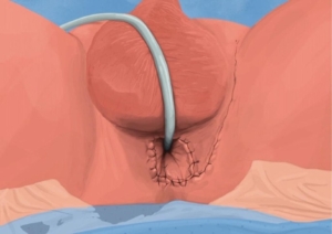
  • An indwelling catheter is inserted, and the bladder is then distended with fluid (saline).
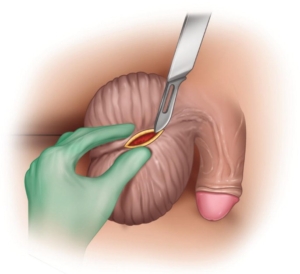
  • A small lower abdominal incision is made, splitting the Linea alba and opening the distended bladder in the midline.
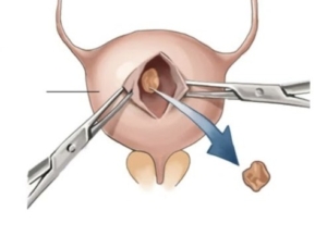
  • The stone is removed through the whole with a grasping instrument.
  • The bladder is inspected and then closed in 2 layers.
  • Skin is closed.
  • A catheter will be left for 2 weeks.

What to expect after the procedure?

  • Hematuria (blood in your urine)
  • You will have an indwelling catheter (IDC), which will remain in your bladder.
  • You may have a continuous bladder irrigation with Saline to help clear the bleeding.
  • When your urine is clear and your bowels are functioning, you will be discharged with catheter care instruction.
  • You will have this indwelling catheter for 2 weeks.
  • A cystogram will be arranged at approx. 14 days to exclude any urine leaks prior to removal of your catheter.
  • If there are any urine leaks, your catheter will remain a further 7 days, or until the leak is sealed.
  • Pain on initial passing of urine when the catheter is removed.
  • Bladder infection ranging from a burning sensation to, fever, to puss (rare).
  • Lower abdominal discomfort which will persist for a few days.
  • NB! Each person is unique and for this reason symptoms vary.

What next?

  • This all depends on what is found during the procedure. All the options will be discussed in detail.
  • There may be some blood in the urine. This can be remedied by drinking plenty of fluids until it clears.
  • Anatomical causes of the stones will be discussed and surgical options in treatment may be discussed
  • Patients should schedule a follow-up appointment within 4-6 weeks to discuss the etiology of the calculus as well as what other procedures may be involved to prevent this from occurring again.


Download Information Sheet

Wes Cysto-Lithotomy Open

Nephro / Uretero Lithotomy – Robotic Assisted

Robotic removal of stones where endoscopic procedures have failed.


Why is it done?

Open/robotic surgery for large or complicated renal and/or ureteric calculi where all other techniques have failed. Seldomly done today. Only if no other options available.

  • As a last resort to remove a large stone.
  • Robotic would be considered first
  • Where equipment is not available.
  • Same entry as for open nephrectomy.

How is it done

  • GA
  • Entry ports for the robot will be determined by where the stone is
  • A ureteric stent would already be in place, if not, this will be attempted prior to robotic access
  • Kidney:
    • Usually an area of thinned cortex
    • Opened and stone removed
    • Closed
  • Renal Pelvis:
    • Opened and stone removed
    • Closed
  • Ureter:
    • Expose the area of the ureter
    • Compression of ureter above and below the calcalculus, as to prevent migration
    • Ureter opened
    • Stone removed
    • Closed
  • Drain left

Alternatives

  • PCNL.
  • ESWL.
  • Sandwich therapy: Combination of PCNL and ESWL.
  • URSE with laser.


Download Information Sheet

Wes Nephro Uretero-lithotomy Open

Optic Urethrotomy

Why is it done?

  • To treat a narrowing in the urethra which has formed due to previous damage/injury to the urethra.
  • Causes:
    • After bypass surgery where a drop in blood pressure has caused an area of low blood supply to the urethra;
    • Trauma to the urethra (pelvic fractures/ urethral instrumentation);
    • Sexually transmitted diseases.
  • The procedure entails cutting the stricture with a cold knife.

How is it done?

  • A urethroscopy is performed by placing a camera in the urethra, with the help of a lubricant jelly and an irrigate fluid, to identify the stricture.
  • A cold knife is then used to cut the stricture open.
  • The inside of the bladder is viewed for pathology.
  • If any suspicious lesions are seen, a biopsy will be taken.
  • Prophylactic antibiotics may be given to prevent infection.
  • Indwelling catheter placed.


Complications

Side–effects

  • Patients will spend the night in the hospital.
  • Patients will be sent home with a catheter for 3 days after receiving thorough catheter care Instructions.
  • Arrangements will be made to remove the catheter on day 3.
  • There may be some blood in the urine. This can be remedied by drinking plenty of fluids until it clears.
  • Review at 6 weeks.
  • There is a >50% risk of recurrence and may need further treatment
    • Re-dilatation
    • Self-dilatation
    • Urethraplasty


Download Information Sheet

Wes Optic Urethrotomy

Copyright 2019 Dr Jo Schoeman

Orchidectomy – Simple

Simple surgical removal of a sick or no functioning or painful testis, with/ without the placement of a prosthesis


Why is it done?

  • To remove a symptomatic non-functioning testis.
  • To remove remnants of a testis after destruction with abscess or infection / TB.
  • To remove a shattered testis after severe trauma.

How is it done?

  • This procedure is performed under general anesthetic.
  • A single incision is made on the midline raphe of the scrotum.
  • The affected testis and vas deference is then extracted through this incision.
  • The testis cord is then exposed as far as possible up in the inguinal area.
  • The blood supply and the vas deferens is separated and tied and cut separately.
  • The cord is tied off twice.
  • The testis is then removed.
  • The cord is checked for hemorrhaging.
  • A drain may be placed
  • A catheter may be left over night.
  • A dressing is then applied, which should be removed after 72 hours.
  • No strenuous movements are permitted for at least 14 days.

NB! You are required to bring 2 pairs of tight new undies for post-operative scrotal support.

What to expect after the procedure?

  • Any anesthetic has its risks, and the anesthetist will explain all such risks.
  • The drain will be removed the next morning.
  • The catheter will be removed 6-8 hours after the procedure.
  • A haematoma (blood collection under the skin or in the scrotal cavity) may form and needs to be reviewed as soon as possible. This may require drainage. Bruising is normal.
  • An infection of the wound can occur and requires immediate attention.
  • Owing to the nature of the surgery and the soft skin of the scrotum, bruising may appear to be much worse than it actually is and is no cause for alarm.
  • DANGER SIGNS: A scrotum that swells immediately to size of a football, fever, puss. Contact Dr Schoeman or the hospital immediately as this may occur in up to 15% of all cases.

What next?

  • The dressing should be kept dry for the initial 72 hours after surgery and then soaked in a bath until the dressing comes off with ease.
  • The dressing may sometimes adhere to the wound causing slight bleeding on removal. Don’t panic, the bleeding will stop.
  • Do not tug at the sutures!
  • PLEASE CONTACT THE HOSPITAL DIRECTLY WITH ANY POST OPERATIVE CONCERNS AND RETURN TO THE HOSPITAL IMMEDIATELY SHOULD THERE BE ANY SIGNS OF SEPSIS.

NB! Regular self-examination highly recommended.

Download Information Sheet

Wes Orchidectomy Simple

Partial Penectomy

Partial amputation of penis with penile block for postoperative pain management.

Why is it done?

  • Confirmed penis cancer, only infiltrating the distal penis.
  • No lymph nodes involved.

How is it done?

  • This procedure is done under general anesthetic.
  • Supine position.
  • The foreskin may be the only affected area and therefore a circumcision is done.
  • Otherwise, the affected area is exposed and cleaned.
  • The affected lesions, usually including the glans is then removed surgically.
  • A 1-2 cm clear surgical margin should be obtained, with sufficient penile length to allow effective urination.
  • The urethral meatus is reconstructed.
  • Hemostatic dressings are placed.
  • Specimen is sent to a histopathologist.
  • An indwelling catheter will be inserted.
  • A dressing is then applied, which should be removed after 72 hours.
  • A local anesthetic is injected at the base of the penis as a penile block thus giving post-operative pain relief for the next 4-6 hours.

What to expect after the procedure?

  • Any anesthetic has its risks, and the anesthetist will explain such risks.
  • Bleeding can occur.
  • Your catheter will be removed on Day 3.
  • Long-term risk of a meatal stenosis.
  • An infection of the wound may occur and requires immediate attention.
  • DANGER SIGNS: A wound that swells immediately, fever, and puss. Contact Dr Schoeman or the hospital immediately as this occurs in up to 15–20% of all cases.

What next?

  • Dressings should be kept dry for the initial 72 hours after surgery and soaked off in a bath thereafter.
  • The dressing may sometimes adhere to the wound causing slight bleeding on removal. Don’t panic, the bleeding will stop.
  • The catheter will be removed after 3 days
  • Patients should schedule a follow-up appointment with Dr Schoeman 4-6 weeks after the procedure.
  • At this stage: if you have any inguinal lymph nodes are palpated, you will be placed on antibiotics. This will be reviewed in 6 weeks.
  • Depending on the staging of the Penile Cancer, Inguinal Node dissection will be scheduled.
  • If your nodes are positive for cancer, Radiation and Chemotherapy will be discussed by an Oncologist.
  • There will be signs of bruising for at least 10 days.
  • Refrain from using your erect penis for 6 weeks.
  • PLEASE CONTACT THE HOSPITAL DIRECTLY WITH ANY POST-OPERATIVE CONCERNS AND RETURN TO THE HOSPITAL IMMEDIATELY SHOULD THERE BE ANY SIGNS OF SEPSIS.


Download Information Sheet

Wes Penectomy Partial

Penile Biopsy

Biopsy of penile skin / lesion to confirm or exclude penile cancer


Why is it done?

  • To confirm/ exclude the presence of malignancy
  • To find the best effective treatment option for this lesion

How is it done?

  • This procedure is done under local or general anesthetic.
  • Supine position.
  • The foreskin may be the only affected area and therefore a circumcision is done.
  • Otherwise, the affected area is exposed and cleaned.
  • If it is a large area, a wedge resection of an area close to normal skin is done.
  • Otherwise, a complete excision biopsy is done.
  • Hemostatic sutures are placed.
  • Hemostatic dressings are placed
  • Specimen is sent to a histopathologist.
  • An in catheter may be inserted if the biopsy area involves the meatus of your urethra.
  • A dressing is then applied, which should be removed after 72 hours.
  • A local anesthetic is injected at the base of the penis as a penile block thus giving post-operative pain relief for the next 4-6 hours.

What to expect after the procedure?

  • Any anesthetic has its risks, and the anesthetist will explain such risks.
  • Bleeding is a common complication.
  • A hematoma (blood collection under the skin). Bruising is normal.
  • Sutures may tear loose with vigorous use of erect penis, and the procedure may then require revision.
  • An infection of the wound may occur and requires immediate attention.
  • DANGER SIGNS: A wound that swells immediately, fever, and puss. Contact Dr Schoeman or the hospital immediately as this occurs in up to 15–20% of all cases.

What next?

  • Dressings should be kept dry for the initial 72 hours after surgery and soaked off in a bath thereafter.
  • The dressing may sometimes adhere to the wound causing slight bleeding on removal. Don’t panic, the bleeding will stop.
  • Patients should schedule a follow-up appointment with Dr Schoeman 2 weeks after the procedure.
  • There will be signs of bruising for at least 10 days.
  • Refrain from using your erect penis for 3-4 weeks.
  • The suture-line will be hard and indurated for at least 8-10 weeks.
  • PLEASE CONTACT THE HOSPITAL DIRECTLY WITH ANY POST-OPERATIVE CONCERNS AND RETURN TO THE HOSPITAL IMMEDIATELY SHOULD THERE BE ANY SIGNS OF SEPSIS.


Download Information Sheet

Wes Penile lesion exision Biopsy

Penile Fracture Repair

Repair of a ruptured Corpora Cavernosa which may sometimes involve a repair of the urethra as well.


How does this occur?

  • Classically seen in guys turning over onto an erect penis in their sleep.
  • Vigorous sex where the erect penis slips out and is re-inserted outside the desired orifice, causing a bend and snap in the corpora cavernosa with a loud snap sound signifying a tear of the CC.
  • Occurs at any age.
  • Sudden severe swelling of penis turning blue, looking like an eggplant.
  • Pain occurs at the time of incident.
  • This requires acute attention, therefore make your way to an emergency department as soon as possible.

What to do?

  • URGENT.
  • Make your way to ED ASAP
  • An ultrasound is done to isolate the tear in the Corpora.

How is it fixed?

  • This fixed under general anesthetic.
  • Supine position (on your back), sterile procedure.
  • An incision is done over the isolated spot.
  • The corpora is sutured with non-dissolving sutures.
  • Where the area is not seen on ultrasound, the foreskin is loosened under the glans with a circumferential incision and the whole penile skin is retracted to the base of the penis.
  • Occasionally a circumcision may result due to complications with this technique, yet foreskin preservation is attempted.
  • An indwelling catheter will be inserted until the swelling is better.
  • A dressing is then applied, which should be removed after 72 hours.

What to expect after the procedure?

  • Any anesthetic has its risks, and the anesthetist will explain such risks.
  • Bleeding is a common complication.
  • A hematoma (blood collection under the skin) is present and will take some time to settle. Bruising is normal.
  • Sutures may tear loose with vigorous use of erect penis, and the procedure may then require revision.
  • An infection of the wound may occur and requires immediate attention.
  • Necrosis of the foreskin can occur in rare circumstances.
  • DANGER SIGNS: A wound that swells immediately, fever, and puss. Contact Dr Schoeman or the hospital immediately as this occurs in up to 15–20% of all cases.

What next?

  • Dressings should be kept dry for the initial 72 hours after surgery and soaked off in a bath thereafter.
  • The dressing may sometimes adhere to the wound causing slight bleeding on removal. Don’t panic, the bleeding will stop.
  • The catheter will be removed as soon as you are awake, or if there are concerns, the following morning.
  • Patients should schedule a follow-up appointment with Dr Schoeman 2 weeks after the procedure.
  • There will be signs of bruising for at least 10 days.
  • Refrain from using your erect penis for 6 weeks.
  • The suture-line will be hard and indurated for at least 8-10 weeks.
  • PLEASE CONTACT THE HOSPITAL DIRECTLY WITH ANY POST-OPERATIVE CONCERNS AND RETURN TO THE HOSPITAL IMMEDIATELY SHOULD THERE BE ANY SIGNS OF SEPSIS.


Download Information Sheet

Wes Penile Fracture Repair

Penile Frenuloplasty

  • The penile tip has a ventral curvature with erection
  • The frenulum can tear and bleed with intercourse


Why is it done?

  • To straighten out a curved penis which is pulled ventrally by a tight frenulum


How is it done?

  • This is done under general anesthetic or a penile block.
  • A horizontal cut is made through the tightest part of the frenulum
  • The incision is then closed by opposing edges in the vertical plane.
  • Dissolvable sutures are placed between the 2 remaining edges.

A local anaesthetic is injected into the base of the penis thus giving postoperative pain relief for the next 4-6 hours

Complications

  • Any anaesthetic has its risks and the anaesthetist will explain such risks.
  • Bleeding is a common complication.
  • With any subsequent erections postoperatively, the sutures may pull out causing an opening of the wound with subsequent bleeding.
  • An infection of the wound can occur

NB! Each person is unique and for this reason, symptoms may vary


Download Information Sheet

Wes Frenuloplasty

Copyright 2019 Dr Jo Schoeman

Penile Shunt / Aspiration

Treatment of a low-flow priapism.


Why is it done?

  • Prolonged painful erections lasting > 4-6 hours.
  • Usually associated with drug use.
  • Usually associated with intracavernosal administration of Erectile Dysfunction drugs.
  • You would have conservative measures, i.e. Icepacks, Pseudo-ephedrine tablets etc.

How is it done?

  • This procedure is done under general anesthetic.
  • Supine position.
  • The penis is surgically prepared.
  • 2 large-bore cannulas are placed through the glans penis into the Corpora Cavernosa.
  • Old clotted blood is drained until flaccid.
  • The corpora is rinsed with saline.
  • A weak mixture of Ephedrine may also be used to rinse the Corpora Cavernosa.
  • Should this not be effective, a scalpel can be placed to cut the CC to create a short circuit with the CS.
  • This may have erectile dysfunction as a result.
  • If the priapism is deflated, the procedure is completed by placing an Indwelling Catheter and an elasticated compression bandage for a few hours.

What to expect after the procedure?

  • Any anesthetic has its risks, and the anesthetist will explain such risks.
  • Bleeding is a possible complication.
  • Your catheter will be removed on Day 2-3.
  • Recurrent priapism requiring more than 1 intervention.
  • The possibility of permanent Erectile Dysfunction.

What next?

  • Dressings should be kept dry for the initial 72 hours after surgery and soaked off in a bath thereafter.
  • The catheter will be removed after 3 days.
  • Patients should schedule a follow-up appointment with Dr Schoeman 4-6 weeks after the procedure.
  • An arteriogram may be recommended to exclude AV-Fistulas.
  • There will be signs of bruising for at least 10 days.
  • PLEASE CONTACT THE HOSPITAL DIRECTLY WITH ANY POST-OPERATIVE CONCERNS AND RETURN TO THE HOSPITAL IMMEDIATELY SHOULD THERE BE ANY SIGNS OF SEPSIS and RETURN OF PRIAPISM.


Download Information Sheet:

Wes Penile Shunt Aspiration

Perineostomy – Perineal Urostomy

Opening of the posterior Urethra on the Perineum

Why is it done?

  • Seldom done
  • This procedure is performed when concentric extensive scarring in the urethra (strictures) causes Urinary Retention
  • A long history of strictures.
  • This is alternative to an invasive procedure where long periods of anesthetic are contra-indicated and extensive grafts may be required
  • Usually for chronically sick patients who cannot undergo surgery yet are active enough not to want a permanent catheter.
  • Patients who don’t want to / cannot do intermittent self-dilatation of these strictures
  • Don’t want a permanent Indwelling Catheter penile/Urethral cripple


How is it done?

  • Patients will receive a General Anesthetic.
  • Flexible cystoscopy is done through your urethra or suprapubic catheter site to find normal urethra (usually posterior urethra)
  • A urethral sound is placed/ catheter
  • A perineal incision is made.
  • The urethra is divided proximal (above) the stricture
  • The opening of the urethra is brought out and attached to the skin on the perineum (the area between scrotum and anus)
  • A catheter is placed
  • Prophylactic antibiotics will be given to prevent any infections.


Complications

  • Persistent pain in penile shaft
  • Pain in Perineum when seated
  • Scarring of the opening requiring dilatation
  • Possible infection
  • NB! Each person is unique and for this reason, symptoms vary!


Download Information Sheet

Wes Perineostomy

Copyright 2019 Dr Jo Schoeman