Pelvi-Ureteric Junction Repair (PUJ) – Robotic Assisted

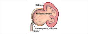
A congenital or acquired narrowing in the ureteric pelvis junction. This narrowing is excised with a reconnection. There are several techniques described in repairing this: I prefer the Dismembered Pyeloplasty

Why is it done?

  • High grade obstruction.
  • Causing deterioration of renal function.
  • Thinning of renal cortex.
  • Chronic pain.
  • Chronic infection.
  • Recurrent renal calculi.

Causes

  • Congenital lack of muscle, or neuro transmission in this area, causing a non-functioning part leading to obstruction.
  • Vesico-ureteric reflux, longstanding can also cause this.
  • Usually diagnosed in kids.
  • Crossing vessel.

How is it done?

Robotic assisted pyeloplasty.

  • Types
      • Dismembered.
      • Foley’s Y-V Pyeloplasty.
      • Culp-Dewierd.
      • Pelvi-calyceal pyeloplasty.
      • Endopyelotomy with laser.
    • Patients will receive a general anaesthesia.
    • Prophylactic antibiotics is given.
    • The correct ureteric system is identified and marked while you are awake.
    • This will be mostly a robotic / laparoscopic procedure.
    • The endoscopic procedure is reserved as a second line in my practice.
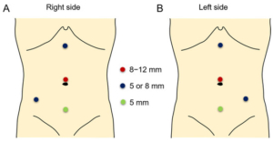
    • Laparoscopic ports are placed
    • The affected ureter is exposed, the defect cut out with a re-anastomosis of a spatulated ureter to a trimmed renal pelvis over a ureteric stent.
    • An indwelling catheter is placed. A drain is placed.

What next?

  • You may be in hospital for 3 days
  • Your drain will be removed when there is no urine draining.
  • Your catheter will be removed the following day.
  • As soon as you are comfortable with no signs of pain and emptying your bladder sufficiently, you will be discharged
  • A ward prescription may be issued on your discharge, for your own collection at any pharmacy.
  • A follow-up appointment will be scheduled for 6 weeks to remove your ureteric stent under local anaesthesia with a Flexible Cystoscope.
  • A review with a CT IVP will be scheduled 6 weeks after this to check on the end result of the ureter.
  • Any pain or signs of fever require an urgent review.
  • DON’T SUFFER IN SILENCE, OR YOU WILL SUFFER ALONE!

Possible Complications

  • Re-stenosis with recurrent obstruction.
  • Second procedure.
  • With further deterioration of renal function, you may require a nephrectomy where affected kidney contributes < 15-20% of total renal function.
  • Urine leak, Urinoma, requiring drainage.
  • Infection possible sepsis requiring long-term antibiotics.


Download Information Sheet
Wes RA reair

Rigid Cystoscopy, Retrograde Pyelogram, Stent Management

A therapeutic procedure under general anaesthetic, where a rigid cystoscopy is done in the bladder via the urethra, ureteric catheters are placed to enable imaging of the upper tracts with/without insertion or removal of ureteric stents

Why is it done?

To investigate:

  • Hematuria (blood in the urine)
  • Recurrent upper urinary tract infections
  • Space occupying lesions in the kidneys and ureters
  • Abnormal cells suggestive of urothelial carcinoma, on urine cytology


Risk factors:

  • Strong family history of bladder cancer
  • Smokers or passive smokers
  • Factory workers: dyes, paints, etc
  • Exposure to Schistosoma (Bilharzia)
  • Renal stone disease, bladder stones


How is it done?

  • This is done under General anaesthesia.
  • A cystoscopy is performed by placing a camera in the urethra with the help of a lubricant gel and saline irrigation.
  • The bladder is then distended with saline.
  • The inside of the bladder is viewed for pathology.
  • A retrograde pyelogram is done at the same time, (placement of small silicone catheters up the kidney pipes). Through this iodine contrast is injected up into the kidney collecting systems. This facilitates the viewing of kidney pipes and kidney collecting systems on X-ray to exclude any upper tract pathology.
  • If any abnormalities are found in the kidney/ ureters, a ureteroscopy (which is the placement of a long thin camera up the ureter) will be performed.
  • If any suspicious lesions are seen, a biopsy will be taken.
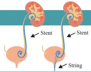
  • A ureteric stent may be placed
  • Urine would have been sent for cytology, to rule out the existence of cancer.

Antibiotics may be given to prevent infection

Complications

What to expect after the procedure?

  • Pain on initial passing of urine
  • Bladder infection ranging from a burning sensation to, fever, to puss (rare)
  • Bloodstained urine
  • Lower abdominal discomfort which will persist for a few days
  • Pain radiating from bladder to renal angle associated with urinating.
  • An infection could present with a stent being present.


Indications for a Ureteric stent

· Hematuria from upper tracts

· Dis-obstruction of the ureter caused either calculus, blood clot or tumour

· External compression of the ureter by retro-peritoneal pathology i.e.: Fibrosis, retroperitoneal lymph node compression

· Reduced renal function associated with hydronephrosis

· Sepsis associated with hydronephrosis


Download Information Sheet

Wes Cystoscopy RGP and Ureteric stents

Copyright 2019 Dr Jo Schoeman

Uretero-Renoscopic Stone Extraction with Laser (URSE)

A ureteric or renal calculus is removed with technique and may require a laser. A rigid/ flexible ureteroscope can be used.

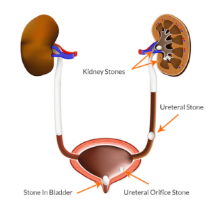
Stones in the kidney, urinary bladder and ureter. medical illustration with a cross section of the kidney and bladder. anatomy of the urinary system. Human kidney.

Why is it done?

  • Removal of renal or ureteric stones.
  • Ureteric stones can vary from 5mm to over 1 cm in size.
  • You may present with excruciating pain on the affected side. (This pain may be worse than childbirth).
  • Renal stones usually larger than 1cm obstructing the renal pelvis.
  • Or renal stones not causing any symptoms.
  • Any fevers or a single kidney is deemed an emergency!

Two Treatment Options

  • Ureteric Calculi.
    • Managed with rigid ureteroscopy.
    • Prior stenting with a ureteric stent,
    • 7-10 days after stenting the stent is removed, and the stone is addressed with laser
  • Renal Calculi.
    • Prior stenting for 7-10 days.
    • After stent removal the kidney is accessed with a flexible uretero-renoscope and the stone is fragmented with laser

How is it done?

    • Patients will receive a general anesthesia.
    • Prophylactic antibiotics is given.
    • The correct kidney is identified and marked while you are awake.
    • You would have had a cystoscopy with retrograde pyelogram 10 days prior with placement of ureteric stent to prepare your ureter.
    • A cystoscopy will be done first to remove the stent, and 2 guidewires will be placed to enable access up the ureter.
    • Depending on the position of the stone, either a rigid or flexible uretero-renoscope will be used.
    • If a stone is in the kidney a flexible uretero-renoscope will be used with access obtained with an access sheath to protect the ureter from damage.
    • Laser will be used to fragment the stone.
    • All fragments will be attempted to be cleared. Small 1-2 mm fragments may be left as “Clinically Insignificant Fragments (CISF)” and will pass spontaneously.
    • A Ureteric catheter with an indwelling catheter is left post-operatively overnight.
    • Catheters will be removed the next morning depending on the presence of blood in the urine.

What next?

  • You will spend at least one night in hospital.
  • You will have a catheter for that time.
  • On removal of your catheter, you may experience sharp colicky pain, exactly the same as your presenting renal colic. This is due to your ureter contracting back to its usual size. (The stent has dilated this to 5X its usual size).
  • You will be discharged as soon as your pain has stabilized and you can function independently.
  • Allow for a few days for stabilization of symptoms.
  • A ward prescription will be issued on your discharge, for your own collection at any pharmacy.
  • A follow-up appointment will be scheduled for 6 weeks. Stone analysis results will then be discussed in order to formulate a plan to proven recurrences
  • DON’T SUFFER IN SILENCE, OR YOU WILL SUFFER ALONE!


Types of Stones:

  • Calcium Oxalate.
  • Uric Acid.
  • Calcium Phosphate.
  • Struvite (Infection stones).
  • Cystine.


Download Information Sheet

Wes Uretero-Renoscopic Stone Extraction with Laser URSE

URSE with or without Laser