UroLift for Benign Prostatic Enlargement: A Minimally Invasive Treatment That Preserves Ejaculation

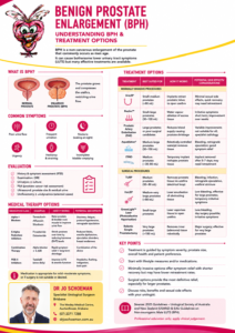
Benign prostatic hyperplasia, or BPH, is a non-cancerous enlargement of the prostate. As the prostate enlarges, its lateral lobes may compress the urethra and obstruct the flow of urine from the bladder.

Common lower urinary tract symptoms include:

  • A weak or interrupted urinary stream
  • Difficulty starting urination
  • Straining to pass urine
  • Urinary frequency and urgency
  • Getting up repeatedly at night
  • Dribbling after urination
  • A feeling that the bladder has not emptied properly

Medication is usually the first treatment offered to men with troublesome symptoms. However, tablets may provide insufficient relief or cause dizziness, tiredness, reduced libido, erectile problems or altered ejaculation.

The UroLift procedure—also called a prostatic urethral lift or PUL—is a minimally invasive surgical therapy, or MIST, that may provide an alternative to long-term medication and conventional prostate surgery.

What is a MIST procedure?

Minimally invasive surgical therapies are designed to improve urinary symptoms with less tissue damage, less bleeding and a shorter recovery than conventional procedures such as TURP or prostate laser surgery.

Different MIST procedures work in different ways. Some use steam or other forms of energy to destroy prostate tissue. Some temporarily remodel the urinary passage. UroLift mechanically moves the obstructing prostate tissue away from the urethra using small permanent implants.

Its principal attractions are:

  • No cutting or removal of prostate tissue
  • No heat, laser or steam
  • Rapid improvement in many patients
  • Usually brief catheterisation, if any
  • Short recovery
  • A low reported risk of new erectile or ejaculatory dysfunction

The trade-off is that symptom improvement is generally more modest than after a tissue-removing operation, permanent implants remain within the prostate, and some patients will eventually require further treatment.

How does UroLift work?

The UroLift system uses small implants to hold the enlarged lateral lobes of the prostate away from the urethra.

Each implant consists of:

  • A small capsular tab placed on the outside of the prostate
  • A stainless-steel urethral end-piece
  • A permanent suture connecting the two components

The implant compresses and retracts the obstructing prostate tissue, widening the urinary channel without cutting, burning or removing tissue.

Most patients require several implants. The exact number depends on the length, size and shape of the prostate and the degree of obstruction. These implants remain permanently in position.

Who may benefit from UroLift?

UroLift may be considered for men who:

  • Have moderate or severe urinary symptoms caused by BPH
  • Have obtained inadequate relief from medication
  • Cannot tolerate the side effects of BPH medication
  • Prefer not to take daily medication indefinitely
  • Want a less invasive alternative to TURP or laser surgery
  • Place a high priority on preserving forward ejaculation
  • Want a relatively rapid return to normal activities
  • Have prostate anatomy suitable for a prostatic urethral lift
  • Understand the possibility of future retreatment

It is particularly attractive for sexually active men who wish to improve their urinary symptoms while minimising the risk of dry or retrograde ejaculation.

What prostate size is suitable?

Clinical guidelines and manufacturer labelling are not identical.

Current European Association of Urology guidance recommends offering prostatic urethral lift to men interested in preserving ejaculation who have prostates smaller than 70 mL and no obstructing middle lobe.

American Urological Association guidance supports the procedure for selected men with prostates approximately 30–80 mL and without an obstructing middle lobe.

Manufacturer and regulatory indications may extend to prostates as large as 100 mL and may include selected median-lobe anatomy in some jurisdictions. However, treatment outside the populations best supported by clinical guidelines requires careful patient selection and counselling.

Prostate volume alone does not determine suitability. The shape of the prostate, length of the prostatic urethra, bladder-neck anatomy, degree of obstruction and strength of the bladder muscle must also be considered.

What about an obstructing median lobe?

The median lobe is prostate tissue that projects upwards towards the bladder and may act like a ball valve over the bladder outlet.

UroLift can be used to treat selected obstructing median lobes, and the MedLift study reported encouraging outcomes. Nevertheless, major guideline recommendations remain more conservative because the strongest long-term randomised evidence relates mainly to lateral-lobe obstruction.

A large, mobile or unusually shaped median lobe may be better treated with TURP, laser surgery, Rezūm, Aquablation or another procedure capable of removing or reducing the obstructing tissue.

This is an important anatomical caveat to discuss before choosing UroLift.

Assessment before treatment

Not every urinary symptom is caused by BPH. Infection, urethral stricture, bladder weakness, overactive bladder, neurological disease, medication and prostate or bladder cancer may produce similar symptoms.

Assessment may include:

  • Medical history and examination
  • International Prostate Symptom Score
  • Urine testing
  • PSA testing when appropriate
  • Urinary-flow measurement
  • Ultrasound measurement of residual urine
  • Prostate imaging or ultrasound
  • Flexible cystoscopy
  • Urodynamic testing in selected patients

Cystoscopy may be particularly useful because it allows the urologist to assess the lateral lobes, bladder neck, median lobe and length of the obstructing prostate.

Any urinary infection should be treated before the procedure.

Patients taking aspirin, warfarin, clopidogrel, apixaban, rivaroxaban or other blood-thinning medication require an individual plan. Do not stop blood-thinning medication without instructions from your prescribing doctor and urologist.

How is the UroLift procedure performed?

UroLift is usually performed as a day procedure under local anaesthetic with sedation or a short general anaesthetic.

A cystoscope and specialised delivery device are passed through the urethra. No external incision is required.

The urologist:

  1. Examines the urethra, prostate and bladder.
  2. Positions the delivery device within the prostatic urethra.
  3. Moves the obstructing prostate tissue away from the urinary channel.
  4. Deploys an implant to hold the tissue in its new position.
  5. Repeats the process at selected points until an adequate channel has been created.
  6. Inspects the bladder outlet and controls any bleeding.

The procedure commonly takes less than an hour, although this varies with prostate anatomy and the number of implants required.

Because no prostate tissue is removed, there is usually no specimen for laboratory examination.

Will I need a catheter?

Many men can pass urine after the procedure and go home without a catheter. A catheter may nevertheless be required if there is:

  • Significant prostate swelling
  • Inability to pass urine
  • Pre-existing urinary retention
  • A weak bladder muscle
  • Bleeding or clot formation
  • A high residual urine volume

When required, the catheter is usually temporary. Patients with chronic retention or poor bladder function have a greater risk of failing an early trial without a catheter.

What should I expect after UroLift?

Temporary urinary irritation is common during the first few days.

You may experience:

  • Burning or stinging when passing urine
  • Urinary urgency and frequency
  • Pelvic, perineal or penile discomfort
  • Light blood in the urine
  • Bladder spasms
  • A temporarily weaker or more irregular stream
  • Increased night-time urination
  • A feeling of incomplete emptying

These symptoms generally improve over several days, although urinary irritation may occasionally persist for a few weeks.

Some men notice an improved stream soon after treatment. For others, the benefit develops over the following two to six weeks as swelling and irritation settle.

Managing pain and urinary discomfort

Discomfort after UroLift is usually mild to moderate.

Management may include:

Paracetamol

Paracetamol is commonly sufficient for mild discomfort. Take it according to the instructions provided by your doctor or the directions on the packet.

Avoid accidentally taking additional paracetamol contained in cold, influenza or combination pain medicines.

Anti-inflammatory medication

Ibuprofen or another anti-inflammatory medicine may help when medically appropriate. These medicines may not be suitable for patients with kidney impairment, stomach ulcers, certain heart conditions, bleeding disorders, anti-inflammatory-sensitive asthma or anticoagulant medication.

Check with your doctor or pharmacist before taking an anti-inflammatory medicine.

Medication for urinary symptoms

An alpha blocker may be continued temporarily to assist urine flow while swelling settles. Medication for bladder spasm or urinary burning may occasionally be prescribed.

Practical measures

It may help to:

  • Maintain normal hydration without forcing excessive fluids
  • Limit coffee, tea, alcohol, fizzy drinks and energy drinks
  • Avoid constipation
  • Avoid heavy lifting and vigorous exercise for several days
  • Take pain relief before discomfort becomes severe

Severe or worsening pain should be reported rather than simply tolerated.

Possible complications

Most side effects are mild and temporary, but complications can occur.

Common temporary effects

These include:

  • Pain or burning during urination
  • Blood in the urine
  • Urinary urgency
  • Increased frequency
  • Pelvic discomfort
  • Temporary urinary leakage associated with urgency

Urinary retention

Some men cannot pass urine after the procedure and require temporary catheterisation. The risk may be greater in patients with high residual urine volumes, chronic retention, severe obstruction or a weak bladder muscle.

Urinary tract infection

Infection may cause worsening burning, cloudy or offensive urine, fever, chills or feeling generally unwell. Antibiotics may be required.

Bleeding

Light haematuria is common. Significant bleeding, clot retention or the need for further intervention is uncommon but possible.

Implant-related problems

Potential implant complications include:

  • Incorrect placement
  • Implant exposure within the bladder
  • Encrustation or stone formation
  • Migration or loosening
  • Persistent pelvic discomfort
  • The need to remove an implant
  • Difficulty or irritation during later prostate surgery

Implants placed too close to or within the bladder are more likely to develop encrustation and may require endoscopic removal.

Persistent symptoms

UroLift treats obstruction but does not correct every cause of lower urinary tract symptoms. Urgency, frequency and nocturia may persist when they are caused by bladder overactivity, excessive night-time urine production, sleep apnoea, fluid intake, diabetes or other medical conditions.

Need for further treatment

Some patients obtain insufficient relief or develop recurrent symptoms as the prostate continues to enlarge. Further treatment may involve medication, additional implants, removal of exposed implants or another procedure such as TURP, GreenLight laser, HoLEP, Rezūm or Aquablation.

When should I seek urgent help?

Contact your urologist or attend an emergency department if you:

  • Cannot pass urine
  • Develop fever, shaking or chills
  • Feel generally unwell or confused
  • Pass large blood clots
  • Have heavy or persistent bright-red bleeding
  • Develop severe or increasing pelvic pain
  • Cannot keep fluids down
  • Experience pain that is not controlled by the recommended treatment

Does UroLift affect erections or ejaculation?

Preserving sexual function is one of the main reasons patients choose UroLift.

The pivotal L.I.F.T. study reported no new sustained erectile or ejaculatory dysfunction during five years of follow-up. Because the bladder neck, prostate tissue and ejaculatory pathways are not routinely cut or heated, the risk of retrograde ejaculation is substantially lower than with conventional TURP and many tissue-removing procedures.

However, no treatment can guarantee unchanged erections, sensation, orgasm or ejaculation in every patient.

Men who already have erectile or ejaculatory difficulties should not assume that UroLift will correct those problems. It is primarily a treatment for urinary obstruction.

How effective is UroLift?

Clinical studies show meaningful average improvement in:

  • Urinary symptom scores
  • Quality of life
  • Peak urinary-flow rate
  • The patient’s perception of urinary function

Symptoms may improve quickly because the urethra is mechanically opened during the procedure.

However, the average improvement in urinary flow and symptoms is generally less than that achieved with TURP or prostate enucleation. UroLift is therefore best understood as a compromise: less invasive treatment and better preservation of ejaculation in exchange for more modest de-obstruction and a greater chance of later retreatment.

How long does UroLift last?

The implants are permanent, but the symptom relief is not necessarily lifelong.

The pivotal L.I.F.T. study demonstrated sustained average improvements for five years. The reported surgical retreatment rate was approximately 13.6% over five years, or roughly 2–3% per year.

Retreatment estimates vary among studies and real-world populations. The need for further treatment may be influenced by:

  • Prostate size and anatomy
  • Median-lobe obstruction
  • Implant position
  • Severity of the original obstruction
  • Bladder function
  • Continued prostate growth
  • The definition of retreatment used in a study

Patients should distinguish between an implant remaining in position and the treatment continuing to control symptoms. A permanent implant does not guarantee permanent symptom relief.

MRI considerations

UroLift implants are classified as MR Conditional, meaning MRI can generally be performed under specified scanner conditions. Patients should tell the radiology service that they have UroLift implants and provide the implant information card whenever possible.

The metallic components can create image artefact, particularly during prostate MRI. This may obscure portions of the prostate and reduce the diagnostic quality of an MRI used to investigate suspected prostate cancer.

This caveat is particularly relevant for:

  • Younger men with a long future need for prostate surveillance
  • Patients with an elevated or rising PSA
  • Men already undergoing prostate cancer monitoring
  • Patients likely to require prostate MRI or targeted biopsy

Appropriate prostate cancer assessment should be completed before UroLift when clinically indicated.

Can prostate surgery still be performed later?

Yes. TURP, laser surgery, HoLEP and other BPH procedures can be performed after UroLift.

However, the surgeon must account for the permanent implants. Clips may be encountered during resection or enucleation, may affect instruments or laser fibres, and may require removal. Some surgeons therefore consider later surgery technically more complex than treatment of an implant-free prostate.

UroLift does not close the door to future treatment, but it does leave permanent material that the future surgeon must manage.

Who should not undergo UroLift?

Manufacturer contraindications include:

  • Active urinary tract infection
  • Current visible or gross haematuria
  • Urinary incontinence caused by an incompetent urinary sphincter
  • A urethral condition that prevents safe insertion of the delivery device
  • A prostate larger than the maximum permitted by the applicable product labelling

UroLift may also be unsuitable or less predictable in men with:

  • Severe chronic urinary retention
  • A poorly contracting bladder
  • Very high residual urine volumes
  • Urethral stricture disease
  • Bladder stones
  • Recurrent urinary infections
  • Significant ongoing bleeding
  • A very high bladder neck
  • A large or unfavourably shaped median lobe
  • A very large prostate
  • Suspected or untreated prostate cancer
  • Symptoms primarily caused by overactive bladder rather than obstruction
  • A need for maximal and durable removal of obstructing tissue

Advantages of UroLift

Potential advantages include:

  • Minimally invasive day procedure
  • No prostate tissue removed
  • No laser, heat or steam
  • Rapid relief in many patients
  • Short recovery
  • Catheter often avoided
  • Low reported risk of new erectile dysfunction
  • Low reported risk of dry or retrograde ejaculation
  • Future BPH treatments remain possible

Limitations and important caveats

Patients should understand that:

  • Several permanent implants remain inside the prostate
  • Symptom and flow improvements are usually less than after TURP or enucleation
  • Retreatment is more common than after tissue-removing surgery
  • Not all median lobes are suitable
  • UroLift does not prevent continued prostate growth
  • Urgency and nocturia may persist if they have a bladder or medical cause
  • The implants may reduce the quality of future prostate MRI
  • Later prostate surgery remains possible but may be technically more complicated
  • Prostate cancer should be appropriately assessed before treatment
  • UroLift does not provide prostate tissue for laboratory examination

Is UroLift the right option for me?

UroLift can be an excellent option for a carefully selected man who wants meaningful improvement in urinary symptoms, a rapid recovery and a low risk of ejaculatory dysfunction.

It may be less suitable for someone with severe obstruction, chronic retention, weak bladder function, unfavourable median-lobe anatomy or a need for the most powerful and durable improvement possible.

The decision should take account of:

  • Prostate size and shape
  • Presence of a median lobe
  • Severity of symptoms and obstruction
  • Bladder strength and residual urine
  • PSA and prostate cancer risk
  • Previous treatment
  • General health and medication
  • Importance of preserving ejaculation
  • Acceptance of permanent implants
  • Willingness to undergo future retreatment

A urological assessment allows UroLift to be compared fairly with medication, iTind, Rezūm, Aquablation, TURP, GreenLight laser, HoLEP and other appropriate options.

This information is intended for general education and does not replace individual medical advice. Treatment suitability, availability and costs vary. Always follow the instructions provided by your treating urologist.

References

  1. European Association of Urology: Management of non-neurogenic male lower urinary tract symptoms.
  2. American Urological Association: BPH clinical guideline.
  3. Roehrborn CG, et al. Five-year results of the prospective randomised controlled prostatic urethral L.I.F.T. study. Canadian Journal of Urology. 2017;24:8802–8813.
  4. Rukstalis D, et al. Prostatic urethral lift for the treatment of an obstructive median lobe: 12-month results of the MedLift study. Prostate Cancer and Prostatic Diseases. 2019;22:411–419.
  5. UroLift manufacturer safety information.
  6. Benidir T, et al. Impact of the UroLift device on prostate magnetic resonance image quality. Journal of Urology. 2023.
If you are troubled by lower urinary tract symptoms but are not yet ready to farewell your forward ejaculation, come and see your Brisbane urologist, Dr Jo, Uro-Jo, to discuss whether UroLift may be an appropriate option for your BPH.

iTind for Benign Prostatic Enlargement: Temporary Treatment Without a Permanent Implant

Benign prostatic hyperplasia—or BPH—is a non-cancerous enlargement of the prostate. As the prostate grows, it may compress the urethra and interfere with emptying the bladder.

Typical symptoms include:

  • A weak or interrupted urinary stream
  • Difficulty starting urination
  • Straining to pass urine
  • Urinary frequency or urgency
  • Getting up repeatedly at night
  • Dribbling after urination
  • A feeling that the bladder has not emptied completely

Medication is often the first treatment. However, tablets do not help every patient and may cause dizziness, tiredness, reduced ejaculation, erectile difficulties or reduced libido. The iTind procedure offers selected men a minimally invasive alternative to long-term medication or conventional prostate surgery.

What is iTind?

The iTind is a small temporary device made from nitinol, a flexible nickel–titanium alloy with “shape memory.”

It is sometimes called a temporary prostatic stent, although it differs from a traditional stent because it is not intended to remain permanently within the prostate. The device is left in place for only five to seven days and is then removed completely.

While in position, three expanding struts apply controlled pressure to specific areas of the prostate and bladder neck. This remodels the prostatic urethra and creates channels through which urine can flow more easily.

The procedure does not:

  • Permanently implant metal within the prostate
  • Remove prostate tissue
  • Use laser, heat or steam
  • Prevent future prostate treatments if symptoms return

Who may benefit from iTind?

iTind may be considered for men who have bothersome urinary symptoms caused by BPH and who:

  • Have obtained insufficient benefit from medication
  • Have developed side effects from BPH tablets
  • Prefer not to take daily medication indefinitely
  • Want a less invasive alternative to TURP or laser surgery
  • Wish to minimise the risk of ejaculatory or erectile side effects
  • Prefer not to have a permanent prostatic implant
  • Have prostate size and anatomy suitable for the device

The procedure is generally best suited to men with moderate-to-severe symptoms and a prostate measuring approximately 25–75 mL, without a significantly obstructing median lobe.

These measurements are a guide rather than a guarantee of suitability. The shape of the prostate, severity of obstruction and function of the bladder are as important as prostate size.

Assessment before treatment

Urinary symptoms are not always caused by an enlarged prostate. Similar problems can result from infection, urethral narrowing, bladder weakness, an overactive bladder, neurological disease, medication or, less commonly, cancer.

Assessment may therefore include:

  • Medical history and examination
  • International Prostate Symptom Score
  • Urine testing
  • PSA testing when appropriate
  • Urinary-flow measurement
  • Ultrasound measurement of residual urine
  • Prostate ultrasound or other imaging
  • Flexible cystoscopy
  • Urodynamic testing in selected patients

Any urinary infection should be treated before the procedure.

Patients taking aspirin, warfarin, clopidogrel, apixaban, rivaroxaban or other blood-thinning medication require an individual management plan. Do not stop blood thinners without instructions from your prescribing doctor and urologist.

How is the iTind procedure performed?

Insertion

The folded iTind device is passed through the urethra using a small cystoscope. It is positioned in the prostatic urethra immediately below the bladder neck and then released.

The procedure is usually performed as day surgery. Depending on the patient and treatment setting, local anaesthetic, intravenous sedation or a short general anaesthetic may be used.

A soft retrieval string remains attached to the device and passes out through the urethra. It is usually secured to the penis until the device is removed.

Most patients can return home on the same day without a urinary catheter. Occasionally, temporary catheterisation is necessary if the patient is unable to pass urine.

The treatment period

The device remains inside the prostate for five to seven days. Its struts gradually apply pressure to the prostate and bladder neck, creating three longitudinal channels within the urinary passage.

Patients remain at home during this treatment period.

Removal

The device must be removed after five to seven days. It is collapsed into a soft catheter and withdrawn in one piece, usually during a brief outpatient visit.

Local anaesthetic gel is often sufficient, although additional pain relief or sedation may be offered when appropriate. No metal or permanent implant remains inside the prostate after removal.

What should I expect while the device is in place?

The five-to-seven-day treatment period is generally the most uncomfortable part of the process. Common temporary symptoms include:

  • Pelvic or perineal pressure
  • Burning or stinging when passing urine
  • Urinary urgency and frequency
  • Discomfort at the tip of the penis
  • Bladder spasms
  • Light bleeding in the urine
  • Interrupted sleep
  • Awareness of the retrieval string
  • A temporarily weak or irregular urinary stream

These symptoms are usually mild to moderate and commonly improve soon after the device is removed.

Patients should generally avoid heavy lifting, vigorous exercise, cycling and sexual activity while the device and retrieval string are in place. Follow the particular instructions provided by your urologist.

The retrieval string should not be pulled, cut, repositioned or removed by the patient.

How can pain and discomfort be managed?

Pain varies considerably between patients. Some men experience only mild pressure, while others find urinary urgency, bladder spasm or discomfort from the retrieval string more troublesome.

A pain-management plan may include the following measures.

Paracetamol

Paracetamol is usually the first choice for mild-to-moderate discomfort. Take it according to your doctor’s instructions or the directions on the packet.

Check cold, influenza and combination pain medicines carefully, as these may also contain paracetamol. Do not exceed the recommended daily dose.

Patients with significant liver disease or heavy alcohol consumption should discuss paracetamol use with their doctor.

Anti-inflammatory medication

An anti-inflammatory medicine such as ibuprofen may help reduce pain and inflammation when medically appropriate.

These medications may not be suitable for patients with:

  • Kidney impairment
  • Stomach ulcers or gastrointestinal bleeding
  • Heart failure or certain cardiovascular conditions
  • Asthma triggered by anti-inflammatory medication
  • Bleeding disorders
  • Anticoagulant or antiplatelet treatment

Check with your urologist, general practitioner or pharmacist before taking an anti-inflammatory medication.

Medication for bladder irritation

An alpha blocker may occasionally be continued or prescribed temporarily to help urine flow. A bladder-calming medication may be considered if urgency or bladder spasms are particularly troublesome.

These medicines are not necessary for every patient and have their own potential side effects.

Practical measures

It may also help to:

  • Maintain normal hydration without forcing excessive fluids
  • Reduce coffee, tea, cola drinks, alcohol and energy drinks
  • Avoid constipation
  • Wear loose, supportive underwear
  • Avoid heavy lifting and strenuous exercise
  • Take pain relief early rather than waiting until discomfort becomes severe

Strong opioid medication is not usually required. It may worsen constipation and, in susceptible patients, contribute to difficulty emptying the bladder.

Severe or increasing pain is not considered something that should simply be tolerated. Contact the treating practice if the discomfort is not controlled by the recommended measures.

What happens after removal?

Burning, urinary urgency and light bleeding may continue briefly after the device is removed, but these symptoms should progressively improve.

Some patients notice an improvement in their stream soon after removal. For others, improvement develops more gradually over several weeks as irritation settles and the remodelled urinary channels stabilise.

Many patients return to normal daily activities within a few days. Your urologist will advise when you may resume strenuous exercise and sexual activity.

Follow-up may include:

  • Review of urinary symptoms
  • Repeat symptom scoring
  • A urinary-flow test
  • Measurement of residual urine
  • Discussion about continuing or stopping BPH medication

Do not stop established prostate medication unless advised to do so.

Side effects and possible complications

Most side effects are temporary and occur while the device is in place. Reported problems include:

  • Burning during urination
  • Urinary frequency and urgency
  • Pelvic discomfort or pain
  • Blood in the urine
  • Temporary difficulty passing urine
  • Urinary tract infection
  • Bladder spasm
  • Movement or incorrect positioning of the device
  • Temporary catheterisation
  • Failure to obtain sufficient symptom relief

Published studies report different complication rates, but most events have been mild and self-limiting. Temporary haematuria, dysuria, urgency and pelvic discomfort are the most frequently reported problems.

Acute urinary retention, urinary infection or significant bleeding are less common but important complications.

When should I seek urgent medical attention?

Contact your urologist or attend an emergency department if you:

  • Cannot pass urine
  • Develop fever, shaking or chills
  • Feel generally unwell or confused
  • Develop severe or increasing pelvic pain
  • Pass large blood clots
  • Have heavy or persistent bright-red bleeding
  • Cannot keep fluids down
  • Notice that the retrieval string or device has moved
  • Develop pain that is not controlled by the agreed treatment plan

Does iTind affect erections or ejaculation?

One of the principal attractions of iTind is its favourable sexual side-effect profile.

Clinical studies have reported preservation of erectile and ejaculatory function in most treated patients. This contrasts with tissue-removing prostate procedures, which may carry a significant risk of retrograde or absent ejaculation.

Nevertheless, no treatment can guarantee that sexual or ejaculatory function will remain unchanged in every patient.

How effective is iTind?

Studies have demonstrated average improvements in:

  • Urinary symptom scores
  • Quality of life
  • Peak urinary-flow rate
  • The patient’s perception of urinary function

The improvement may be less dramatic than that achieved with a tissue-removing operation such as TURP or laser enucleation. The advantage is that iTind is less invasive, usually requires little recovery time and has a low reported risk of sexual side effects.

It is therefore best viewed as a balance between symptom improvement and treatment invasiveness.

How long does the benefit last?

Although the implant remains in place for only five to seven days, the channels produced within the prostate may remain open for several years.

Prospective studies have demonstrated sustained benefit for more than four years in some appropriately selected patients. This does not mean that iTind is guaranteed to last for four years—or that it will provide lifelong relief.

BPH may continue to progress as a man ages. Some patients will eventually need to restart medication or undergo another procedure. Longer-term evidence for iTind remains more limited than that available for established procedures such as TURP or laser enucleation.

Because nothing is left permanently inside the prostate, iTind does not generally prevent later treatment. Options may include medication, another minimally invasive procedure, TURP, GreenLight laser treatment, HoLEP or another form of prostate surgery.

Who should not undergo iTind?

Contraindications described in the manufacturer’s information include:

  • Active urinary tract infection
  • Acute prostatitis
  • Known prostate cancer
  • Known bladder cancer
  • An artificial urinary sphincter or another implant within the urethra
  • Bladder atonia or a non-contractile bladder
  • Neurogenic bladder dysfunction
  • Urinary obstruction caused by a urethral stricture or something other than BPH

The procedure may also be unsuitable or less predictable in men with:

  • A significantly obstructing median prostatic lobe
  • A prostate outside the best-studied size range
  • Severe chronic urinary retention
  • Very high residual urine volumes
  • Recurrent urinary infections
  • Bladder stones
  • Significant ongoing haematuria
  • Advanced bladder weakness
  • A need for rapid and substantial removal of obstructing tissue
  • Anatomy that prevents safe cystoscopic access

Previous prostate surgery, urethral surgery, nickel sensitivity and blood-thinning medication require individual consideration.

Patients with obstruction causing kidney impairment, repeated retention, recurrent infections, bladder stones or significant bleeding may receive more reliable relief from a definitive tissue-removing procedure.

Advantages of iTind

Potential advantages include:

  • Minimally invasive day procedure
  • No cutting, laser, steam or thermal energy
  • No permanent implant
  • Usually no postoperative catheter
  • Rapid return to ordinary activities
  • Low reported risk of new erectile dysfunction
  • Low reported risk of ejaculatory dysfunction
  • Does not usually compromise future BPH treatment

Limitations of iTind

Potential limitations include:

  • Discomfort during the treatment week
  • A retrieval string for five to seven days
  • A second procedure to remove the device
  • Not suitable for every prostate shape or size
  • Less improvement than some tissue-removing procedures
  • Possibility of persistent or recurrent symptoms
  • Potential need for future medication or surgery
  • Less very-long-term evidence than TURP or laser enucleation

Is iTind the right treatment for me?

iTind is not simply a smaller version of TURP. It offers a different compromise: a less invasive procedure with no permanent implant and a favourable sexual side-effect profile, but generally more modest improvement and less long-term evidence than tissue-removing surgery.

The best treatment depends on:

  • The severity of your symptoms
  • The size and shape of your prostate
  • The amount of urinary obstruction
  • Bladder strength and residual urine
  • Previous treatment
  • General health and medication
  • The importance of preserving ejaculation
  • Your willingness to accept possible future retreatment

A urological assessment is essential to confirm that BPH is responsible for the symptoms and to compare iTind fairly with medication, Rezūm, UroLift, TURP, GreenLight laser, HoLEP and other appropriate treatments.

This information is intended for general education and does not replace individual medical advice. Treatment suitability, availability and costs vary. Always follow the instructions provided by your treating urologist.

References

  1. Sandhu JS, et al. Management of Lower Urinary Tract Symptoms Attributed to Benign Prostatic Hyperplasia: AUA Guideline Amendment 2023. Journal of Urology. 2024.
  2. Chughtai B, et al. The iTind Temporarily Implanted Nitinol Device for the Treatment of Lower Urinary Tract Symptoms Secondary to Benign Prostatic Hyperplasia: A Multicentre, Randomised, Controlled Trial. Urology. 2021;153:270–276.
  3. Kadner G, et al. Second generation of temporary implantable nitinol device in men with lower urinary tract symptoms: two-year results of the MT-02 study. World Journal of Urology. 2020;38:3235–3244.
  4. Amparore D, et al. Three-year results following treatment with the second-generation temporary implantable nitinol device. Prostate Cancer and Prostatic Diseases. 2021;24:349–357.
  5. Amparore D, et al. Temporary implantable nitinol device for BPH-related lower urinary tract symptoms: over 48-month results. Minerva Urology and Nephrology. 2023;75:743–751.
  6. NICE: iTind for lower urinary tract symptoms caused by BPH.
  7. Olympus: iTind clinical and procedural information.

So, if you are experiencing LUTS, lower urinary tract symptoms, and you are not yet ready to farewell your prograde ejaculation function, come see your Brisbane urologist, Dr Jo, Uro-Jo and discuss this option for your BPH.

Barrigel® Rectal Spacer During Prostate Cancer Radiation Therapy

Creating a temporary safety zone between the prostate and rectum

Radiation therapy is an effective treatment for many men with prostate cancer. Modern radiation techniques are remarkably accurate, but there is one anatomical challenge: the prostate sits immediately in front of the rectum.

This means that while radiation is directed at the prostate, part of the rectum may also receive radiation.

One approach to reducing this exposure is to temporarily create a small space between the prostate and rectum before radiation treatment begins.

Barrigel® is a biodegradable hyaluronic acid rectal spacer designed specifically for this purpose.


Why does the rectum need protection?

The prostate and rectum normally sit almost against each other, separated by only a thin layer of tissue.

During prostate radiation therapy, some radiation therefore inevitably reaches the anterior wall of the rectum.

This can contribute to bowel symptoms such as:

  • increased bowel frequency
  • urgency
  • loose stools or diarrhoea
  • rectal discomfort
  • mucus discharge
  • rectal bleeding
  • radiation proctitis

Most bowel symptoms following modern radiation therapy are mild and temporary, but occasionally they can persist or become troublesome.

The basic idea behind a rectal spacer is delightfully simple:

Prostate → spacer → rectum

Instead of asking radiation technology to perform an anatomical magic trick, we physically move the rectum a little farther away.

Even approximately 1 cm of additional separation can substantially reduce the radiation dose received by the anterior rectal wall.


What is Barrigel?

Barrigel is a sterile, biodegradable gel made from stabilised, non-animal-derived hyaluronic acid.

Hyaluronic acid occurs naturally in many tissues within the human body and is widely used in medical applications.

Barrigel is injected into the tissue plane between the prostate and anterior rectal wall before prostate radiation therapy.

The gel creates a temporary cushion that pushes the rectum away from the prostate.

Importantly, Barrigel does not treat the prostate cancer itself.

Its purpose is to protect surrounding normal tissue while radiation treats the cancer.


How is Barrigel inserted?

Barrigel is usually inserted before radiation planning.

The procedure is performed using ultrasound guidance.

A needle is passed through the skin of the perineum, the area between the scrotum and anus, rather than through the rectum.

The needle is carefully positioned between the back of the prostate and the front wall of the rectum.

Barrigel is then slowly injected while the position of the gel is continuously monitored with ultrasound.

Typically several millilitres of gel are used to create the required separation.

The procedure may be performed using:

  • local anaesthetic
  • sedation
  • or another form of anaesthesia depending upon the patient and treating centre.

Fiducial markers used for radiation targeting may sometimes be inserted during the same procedure.

Antibiotic prophylaxis and strict sterile technique are important.


Who may benefit from Barrigel?

Barrigel may be considered for men undergoing radiation therapy for localised prostate cancer where reducing radiation exposure to the rectum is desirable.

This may include men undergoing:

Conventional external-beam radiation therapy

A spacer may reduce the amount of radiation reaching the rectum during a multi-week course of treatment.

Hypofractionated radiation therapy

Modern prostate radiation is increasingly delivered using larger doses over fewer treatment sessions.

A randomised clinical trial involving 201 men demonstrated that a hyaluronic-acid spacer significantly reduced rectal radiation exposure during hypofractionated prostate radiotherapy.

Acute grade 2 or greater gastrointestinal side effects occurred in approximately 2.9% of spacer-treated patients compared with 13.8% without a spacer.

Stereotactic body radiation therapy – SBRT

SBRT delivers relatively high radiation doses over only a few treatments. Precise protection of surrounding structures therefore becomes particularly important.

Selected salvage radiation situations

Spacer placement may occasionally be considered in more complex circumstances, although previous prostate or pelvic treatment can alter the normal tissue planes and make insertion more difficult.

These cases require individual assessment by the radiation oncologist and urologist.


When should Barrigel not be used?

Barrigel is contraindicated in patients with clinical T4 prostate cancer.

It should also be approached cautiously in men with conditions that may make insertion difficult or increase the risk of complications.

These include:

  • active infection or inflammation near the injection site
  • bleeding disorders
  • anticoagulant or thrombolytic therapy
  • significant antiplatelet medication
  • known allergy to hyaluronic-acid products
  • significant anorectal stenosis or anatomical abnormalities
  • previous surgery causing extensive scarring around the prostate or rectum
  • significant haemorrhoidal disease
  • immunodeficiency or significant immunosuppressive therapy.

Anticoagulant and antiplatelet medications should not simply be stopped without medical advice. Their management needs to be individualised according to the reason they are being taken.


What are the potential side effects?

Most patients tolerate spacer insertion well.

Temporary symptoms may include:

  • discomfort in the perineum
  • minor bleeding or bruising
  • a sensation of rectal fullness
  • temporary discomfort when opening the bowels
  • urinary frequency or discomfort
  • weak urinary stream
  • temporary constipation.

The feeling that there is “something there” behind the prostate can occur initially and generally settles.


What are the uncommon but important complications?

Although rectal spacer insertion is generally considered a low-risk procedure, it is still an invasive procedure.

Possible complications include:

  • infection
  • prostatitis
  • bleeding or haematoma
  • urinary retention
  • significant rectal pain
  • difficult or painful defaecation
  • incorrect positioning of the spacer
  • injection into the prostate
  • injury to the urethra or bladder
  • rectal-wall injury
  • rectal ulceration or tissue necrosis
  • vascular injection or embolisation
  • very rarely, fistula formation or severe pelvic infection.

This is why spacer insertion should be performed by clinicians experienced in transperineal ultrasound-guided procedures.

Correct placement matters just as much as the choice of spacer.


What happens to Barrigel afterwards?

Barrigel is biodegradable.

It remains between the prostate and rectum during the period when radiation treatment is being delivered and is subsequently broken down and absorbed by the body.

No second operation is normally required to remove it.


Barrigel versus SpaceOAR®

Barrigel and SpaceOAR are designed to solve the same anatomical problem, but they use different materials.

Barrigel SpaceOAR
Material Stabilised hyaluronic acid Polyethylene glycol hydrogel
Main purpose Separate prostate and rectum Separate prostate and rectum
Placement Transperineal injection Transperineal injection
Imaging guidance Ultrasound Ultrasound
Biodegradable Yes Yes
Remains permanently No No
Material characteristics Hyaluronic-acid gel that can be progressively shaped during injection PEG hydrogel formed after injection
Radiopaque version No equivalent to SpaceOAR Vue SpaceOAR Vue contains iodine for CT visibility
Evidence Randomised evidence demonstrating improved rectal dosimetry and reduced acute GI toxicity Extensive clinical experience and randomised/prospective evidence supporting rectal dose reduction

Both therefore create a temporary physical separation between the prostate and rectum.


Is Barrigel better than SpaceOAR?

At present it would be too simplistic to say that one spacer is universally “better”.

Both can substantially reduce radiation exposure to the rectum when correctly placed.

There are, however, some practical differences.

Barrigel can be shaped during insertion

Barrigel is injected as hyaluronic-acid gel. The operator can progressively deposit and shape the material while watching the developing space with ultrasound.

This can be useful when trying to achieve an appropriate distribution behind the prostate.

SpaceOAR has a long clinical track record

SpaceOAR has been extensively studied and is widely used internationally.

SpaceOAR Vue also contains iodine, making the spacer readily visible on CT. This may be helpful when MRI is unsuitable or when CT-based radiation planning is required.

Placement may matter more than the label on the syringe

An important point is that the quality, volume and position of the spacer can significantly influence the radiation dose received by the rectum.

A 2025 real-world comparative study found differences in dosimetry between Barrigel and SpaceOAR that were strongly influenced by the amount of spacer inserted. At comparable volumes, Barrigel performed favourably for some dosimetric measurements.

Interestingly, rectal-wall infiltration occurred in 7 of 287 SpaceOAR procedures (2.44%) and none of the Barrigel procedures in that particular study.

This does not prove that Barrigel is universally safer, but it highlights the importance of spacer material, injection technique and careful positioning.


What if Barrigel is not perfectly positioned?

One potentially useful characteristic of hyaluronic acid is that it can potentially be treated with hyaluronidase, an enzyme that breaks down hyaluronic acid.

This provides a theoretical and practical advantage if significant malposition is recognised.

The Queensland Clinical Excellence guidance specifically notes Barrigel’s potential to be hydrolysed in situations of spacer misplacement.

This does not make incorrect placement harmless, however. Prevention through meticulous ultrasound-guided insertion remains far preferable to correction afterwards.


Do all men having prostate radiation need a spacer?

No.

Modern radiation techniques such as IMRT, VMAT and image-guided radiotherapy already provide highly sophisticated targeting.

Whether a spacer provides meaningful additional benefit depends upon:

  • prostate anatomy and size
  • distance between prostate and rectum
  • radiation technique
  • radiation dose and fractionation
  • previous prostate or pelvic treatment
  • bowel disease
  • anticoagulation
  • individual risk of radiation toxicity
  • and the experience of the treating radiation team.

Some patients may gain considerable benefit while others may gain relatively little.

The decision should therefore be made jointly between the patient, radiation oncologist and urologist.


What does the evidence tell us?

The European Association of Urology recognises biodegradable rectal spacers as a method of increasing the distance between the prostate and rectum and reducing rectal radiation exposure.

Evidence across rectal spacer studies suggests reductions in both acute and late grade 2 or greater rectal toxicity.

For Barrigel specifically, a multicentre randomised trial demonstrated impressive dosimetric results during hypofractionated radiotherapy.

98.5% of patients receiving the hyaluronic-acid spacer achieved at least a 25% reduction in the volume of rectum receiving 54 Gy.

The average reduction was approximately 85%.

The same study demonstrated substantially fewer acute grade 2 or greater gastrointestinal side effects in patients receiving the spacer.

These findings support rectal spacing as a useful tool for appropriately selected men receiving prostate radiation therapy.


The bottom line

Barrigel is a temporary hyaluronic-acid rectal spacer used to create additional distance between the prostate and rectum before prostate cancer radiation therapy.

It does not improve the radiation by attacking the cancer. Instead, it gives the radiation oncologist a little more anatomical breathing room.

For appropriately selected patients it can:

increase prostate-to-rectum separation → reduce rectal radiation dose → reduce the risk of bowel toxicity.

Barrigel and SpaceOAR both achieve this objective using different biomaterials. Neither should automatically be regarded as the best option for every patient.

The choice should take into account the planned radiation technique, individual anatomy, previous treatment, medical history and the experience of the treating team.

Most importantly, a rectal spacer is an additional protective tool, not a guarantee against radiation-related bowel complications.

When should I discuss a rectal spacer?

If you are considering external-beam radiation therapy or SBRT for prostate cancer, it is reasonable to ask your radiation oncologist or urologist:

“Would I benefit from a rectal spacer, and would Barrigel or SpaceOAR be more appropriate for me?”

That conversation should occur before radiation planning begins.

This information is intended for patient education and does not replace individual assessment by your urologist or radiation oncologist.

So, if you have discussed this with your radiation oncologist and you would like the benefit of protecting your rectum against the effects of radiation, come see your local Brisbane urologist, Dr Jo, to discuss this option.

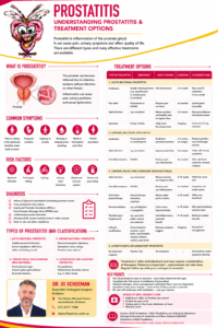
Information Sheet: Prostatitis

Robotic-Assisted Radical Prostatectomy: When Is a Non-Nerve-Sparing Procedure Necessary?

Removing the prostate while putting cancer control first

Robotic-assisted radical prostatectomy is a well-established surgical treatment for localised and selected locally advanced prostate cancer. During the operation, the prostate gland and seminal vesicles are removed, and the bladder is reconnected to the urethra. In selected patients, pelvic lymph nodes may also be removed.

One of the important decisions made before and during prostate cancer surgery is whether the nerves responsible for erections can safely be preserved.

This is known as nerve-sparing prostatectomy.

Unfortunately, nerve preservation is not always appropriate. When prostate cancer is close to, involves, or is suspected of extending beyond the outer edge of the prostate near these nerves, attempting to preserve them may compromise the completeness of cancer removal.

In this situation, a partial or complete non-nerve-sparing robotic prostatectomy may be recommended.

The guiding principle is simple:

Preserve the nerves when it is oncologically safe to do so, but do not preserve them at the expense of adequately treating the prostate cancer.


What are the nerves that are being “spared”?

Running immediately alongside the prostate are delicate bundles of nerves and blood vessels known collectively as the neurovascular bundles.

These structures contain nerves that play an important role in producing erections.

They sit extremely close to the outer surface, or capsule, of the prostate. This anatomical relationship creates a challenge during prostate cancer surgery.

If the cancer is safely contained within the prostate and sufficiently distant from the neurovascular bundle, the surgeon may be able to carefully dissect the prostate away while preserving the nerves.

This is a nerve-sparing radical prostatectomy.

If cancer is suspected to extend towards or through the prostate capsule in this area, the surgeon may need to remove some or all of the neurovascular tissue together with the prostate.

This is a non-nerve-sparing prostatectomy.


Why would a surgeon deliberately remove the erectile nerves?

It may initially seem counterintuitive to remove structures that are so important for sexual function.

The reason is cancer control.

The primary purpose of radical prostatectomy is to completely remove the prostate cancer.

If a tumour is growing very close to the edge of the prostate, particularly next to a neurovascular bundle, dissecting too close to the prostate in an attempt to preserve the nerves could potentially leave cancer cells behind.

This is called a positive surgical margin.

In appropriately selected patients, taking a wider margin around the prostate may therefore provide a safer cancer operation.

The decision represents a balance between:

Cancer control

and

preservation of erectile function.

When these two goals conflict, cancer control generally takes priority.


When may a non-nerve-sparing prostatectomy be recommended?

Non-nerve-sparing surgery may be considered when there is a significant risk that prostate cancer has extended towards or into the tissues surrounding the prostate.

Examples include:

Locally advanced prostate cancer

A tumour suspected of extending through the prostate capsule, particularly clinical T3 disease, may require a wider surgical excision.

Cancer close to the neurovascular bundle on MRI

Modern multiparametric prostate MRI can provide valuable information about the location of the tumour and its relationship to the prostate capsule and neurovascular bundles.

Features suggesting extraprostatic extension may influence the decision not to preserve the nerve bundle on that side.

High-grade prostate cancer

Higher-grade cancers, including cancers with an unfavourable Gleason score or ISUP Grade Group, may have a greater likelihood of extending outside the prostate.

The biopsy result alone does not automatically determine whether the nerves can be preserved, but it forms part of the overall assessment.

Extensive cancer on prostate biopsy

Large-volume disease, particularly when concentrated along the outer portion of the prostate near a neurovascular bundle, may make nerve preservation less appropriate.

Very high PSA or other high-risk features

PSA level, PSA density, biopsy findings, MRI appearance and clinical examination are considered together when estimating the likelihood of disease extending beyond the prostate.

Cancer involving one particular side of the prostate

Importantly, nerve sparing does not necessarily have to be “all or nothing.”

If the cancer is predominantly on one side, it may sometimes be possible to preserve the neurovascular bundle on the opposite side.

This is known as unilateral nerve sparing.


Nerve sparing is not simply YES or NO

Modern robotic prostate surgery is more nuanced than dividing operations into completely nerve-sparing and completely non-nerve-sparing procedures.

Depending on the location and extent of the cancer, surgery may involve:

  • Bilateral nerve sparing
  • Unilateral nerve sparing
  • Partial nerve sparing
  • Wider excision on one side and nerve preservation on the other
  • Complete bilateral non-nerve-sparing surgery

The surgical plan can therefore be tailored to the individual patient and, importantly, to the cancer on each side of the prostate.


How does nerve-sparing surgery differ from non-nerve-sparing surgery?

During nerve-sparing surgery, the surgeon carefully separates the neurovascular bundle from the surface of the prostate while attempting to minimise traction, heat and other potential injury to these delicate nerves.

During non-nerve-sparing surgery, the dissection is deliberately performed further away from the prostate.

The neurovascular tissue is removed together with the prostate where necessary to obtain a wider cancer margin.

The robotic platform provides magnified three-dimensional vision and highly controlled instrument movement. This allows the surgeon to identify tissue planes with considerable precision.

However, robotic technology cannot make an unsafe nerve-sparing operation safe.

The biology and location of the cancer ultimately determine how close to the prostate the surgeon can safely operate.


What happens to erections after non-nerve-sparing prostatectomy?

This is one of the most important issues to discuss before surgery.

The nerves surrounding the prostate are responsible for signalling increased blood flow into the penis to produce a natural erection.

If both neurovascular bundles are completely removed, the likelihood of recovering spontaneous erections adequate for sexual intercourse is very low.

This is different from nerve-sparing surgery, where the nerves remain anatomically intact but may temporarily function poorly following surgery.

After nerve-sparing prostatectomy, erectile recovery can occur gradually over many months and sometimes over 18–24 months or longer.

After complete bilateral non-nerve-sparing surgery, however, the nerves themselves have been removed rather than temporarily stunned.

The expectations for recovery are therefore very different.


Does non-nerve-sparing surgery affect sensation or orgasm?

This is an important distinction.

The nerves responsible for penile sensation are different from the neurovascular nerves primarily responsible for erections.

Many men can therefore continue to experience penile sensation and sexual pleasure following radical prostatectomy.

Orgasm may also remain possible.

However, radical prostatectomy removes the prostate and seminal vesicles and disconnects the reproductive pathway. Consequently, there is no ejaculation of semen after surgery.

This is sometimes described as a dry orgasm.

The sensation of orgasm can also feel different after prostate surgery.


Does non-nerve-sparing prostatectomy cause infertility?

Yes.

Radical prostatectomy results in permanent infertility because the prostate and seminal vesicles are removed and sperm can no longer enter the ejaculate.

Men who may wish to father children in the future should discuss sperm banking before surgery.

This applies whether the operation is nerve sparing or non-nerve sparing.


Is a non-nerve-sparing procedure reversible?

No.

Once a neurovascular bundle has been surgically removed, it cannot simply be reattached at a later date.

This is an important difference between temporary nerve dysfunction and actual nerve removal.

After nerve-sparing surgery, the nerves may be anatomically preserved but temporarily injured or “stunned”. Recovery may therefore occur with time.

After complete non-nerve-sparing surgery, the erectile nerves have been physically removed as part of the cancer operation.

The procedure itself is therefore not reversible.

However, this does not mean that erections or sexual activity are impossible.

There are several effective treatments available for erectile dysfunction following prostatectomy.


Can erections still be achieved after non-nerve-sparing surgery?

Rarely yes, but they will usually require assistance.

Treatment options include:

PDE5 inhibitor tablets

Medications such as sildenafil or tadalafil are commonly used after prostate surgery.

They rely substantially on functioning nerve pathways and are therefore generally more effective following nerve-sparing surgery.

Their effectiveness following complete bilateral non-nerve-sparing surgery is considerably more limited.

Vacuum erection device

A vacuum erection device creates negative pressure around the penis, drawing blood into the erectile tissues.

A constriction ring can then be placed around the base of the penis to maintain the erection.

Because this technique does not depend on intact erectile nerves, it can be useful after non-nerve-sparing surgery.

Penile injection therapy

Medication can be injected directly into the erectile tissue of the penis.

These medications act directly on penile blood vessels and therefore do not require normal prostate-associated nerve signalling.

For this reason, penile injections can be highly effective even after bilateral non-nerve-sparing prostatectomy.

Penile prosthesis

For men with persistent erectile dysfunction who wish to restore reliable erections, a penile prosthesis can provide an effective long-term solution.

An inflatable penile prosthesis allows an erection to be mechanically produced when desired.

For appropriately selected men, satisfaction rates following penile prosthesis surgery are generally high.


What about penile rehabilitation?

Penile rehabilitation may be discussed following radical prostatectomy.

The objectives can include maintaining penile tissue health, encouraging regular oxygenation of the erectile tissues, minimising shortening and fibrosis, and assisting the return to sexual activity.

A rehabilitation program may involve:

  • PDE5 inhibitor medication where appropriate
  • Vacuum erection therapy
  • Penile injection therapy
  • Regular sexual stimulation
  • Early assessment and management of erectile dysfunction

The appropriate program depends heavily on whether surgery was bilateral nerve sparing, unilateral nerve sparing or completely non-nerve sparing.

It is therefore important that expectations are realistic.

After complete bilateral nerve removal, rehabilitation cannot make the removed nerves grow back. Instead, treatment focuses on maintaining penile health and providing alternative ways of achieving an erection.


Can the surgeon decide during the operation whether to spare the nerves?

Sometimes.

The intended degree of nerve sparing is usually planned before surgery using information from:

  • Prostate MRI
  • Prostate biopsy
  • PSA
  • Clinical examination
  • Location and volume of cancer
  • Gleason score and ISUP Grade Group
  • Estimated risk of extraprostatic extension
  • The patient’s existing erectile function
  • The patient’s priorities regarding cancer control and sexual function

However, the final surgical approach may occasionally need to be modified according to findings encountered during the operation.

The most important objective remains adequate removal of the cancer.


Does non-nerve-sparing surgery improve cancer cure rates?

Not every patient benefits from wider surgery.

For men with cancer safely confined within the prostate, unnecessary removal of the neurovascular bundles may produce significant functional consequences without providing additional cancer benefit.

Conversely, when cancer is suspected of extending close to or beyond the prostate capsule adjacent to a neurovascular bundle, wider excision may reduce the risk of leaving tumour at the surgical margin.

This is why the decision must be individualised.

The best operation is not automatically the operation that preserves the most nerves. It is the operation that provides appropriate cancer clearance while preserving as much normal function as can safely be preserved.


What about urinary continence?

Nerve sparing primarily relates to erectile function, rather than the urinary sphincter responsible for continence.

Urinary control after radical prostatectomy depends on several factors including:

  • Age
  • Pre-operative urinary function
  • Pelvic floor strength
  • Urethral length
  • Bladder function
  • Surgical anatomy and technique
  • Previous prostate treatments
  • Individual healing

Pelvic floor rehabilitation before and after surgery can be an important part of recovery.

Non-nerve-sparing surgery does not automatically mean that a patient will remain incontinent.


Questions worth asking before surgery

If a non-nerve-sparing robotic prostatectomy has been recommended, useful questions to discuss with your urologist include:

Why is nerve preservation considered unsafe in my particular cancer?

Is the concern on one side or both sides of the prostate?

Could unilateral or partial nerve sparing be considered?

What does my MRI show about the relationship between the cancer and neurovascular bundles?

What is my estimated chance of erectile recovery with the proposed operation?

What erectile rehabilitation options will be available after surgery?

Should I consider sperm banking before treatment?

These conversations are particularly important because the decision to remove a neurovascular bundle is generally irreversible.


The important message

A non-nerve-sparing robotic-assisted radical prostatectomy is not an inferior version of nerve-sparing surgery.

In the right patient, it is a deliberate cancer-control strategy.

When prostate cancer is close to or suspected of involving the tissues surrounding the prostate, preserving the erectile nerves too aggressively may risk leaving cancer behind.

Modern robotic surgery allows the operation to be tailored to the individual patient. Some men can undergo bilateral nerve preservation, others may benefit from preservation on only one side, while patients with more extensive disease may require a wider non-nerve-sparing excision.

The consequences for erectile function are important and should be understood before surgery.

Once the neurovascular nerves have been removed, the procedure cannot be reversed.

However, loss of spontaneous erections does not mean the end of sexual intimacy. Vacuum devices, penile injection therapy and penile prostheses can provide effective options when natural erections are no longer possible.

Ultimately, the aim is to achieve the best possible balance between two important goals:

Effective treatment of the prostate cancer and preservation of quality of life.


A note for patients

Every prostate cancer is different. MRI findings, biopsy results, PSA, cancer grade, age, general health, pre-existing erectile function and personal priorities all influence the appropriate surgical approach.

A detailed discussion with your urologist before robotic prostatectomy is essential so that you understand whether nerve sparing is appropriate, what degree of nerve preservation may be possible and what this means for cancer control, continence and sexual function.

This information is intended for general patient education and does not replace individual medical advice. Treatment recommendations should be based on your individual prostate cancer characteristics, imaging, pathology, general health and discussion with your treating urologist.

So., come chat to your Brisbane based urologist, Jo to discuss this option with you.

Single-Port Robotic Surgery: The Next Evolution of da Vinci Surgery in Urology

Robotic surgery has transformed modern urology. Now, the technology is evolving again.

The da Vinci SP® (Single Port) Surgical System represents a new generation of robotic-assisted surgery designed to allow complex operations to be performed through one small surgical access point, rather than the multiple abdominal ports traditionally required for robotic surgery.

For suitable patients, this offers an exciting possibility: maintaining the precision and control associated with robotic surgery while potentially reducing the physical footprint of the operation.

What is the da Vinci SP system?

Traditional multi-port robotic surgery generally requires several small abdominal incisions. Each incision accommodates a camera, robotic instrument or assistant port.

The da Vinci SP system takes a different approach.

Through a single approximately 2.7 cm cannula, the system can deploy:

  • Three fully wristed robotic instruments
  • A flexible, articulating 3D high-definition camera
  • Instruments capable of triangulating once inside the body

The surgeon remains completely in control of the operation from the robotic console. The robot does not perform the surgery independently. Rather, it translates the surgeon’s hand movements into precise movements of the instruments inside the patient.

The system provides 360-degree anatomical access and was specifically designed to facilitate surgery within confined anatomical spaces.

Why is Single-Port surgery particularly interesting in urology?

Much of urological surgery occurs deep within relatively confined areas of the body, particularly surgery involving the prostate, bladder and kidney.

This makes urology particularly well suited to the development of single-port techniques.

The da Vinci SP platform is being used internationally for procedures including:

  • Robotic radical prostatectomy for prostate cancer
  • Robotic simple prostatectomy or prostate enucleation for very large benign prostates
  • Partial nephrectomy for selected kidney tumours
  • Radical nephrectomy
  • Pyeloplasty for pelvi-ureteric junction obstruction
  • Selected reconstructive urological procedures

Importantly, the technology also allows surgeons to explore different routes to the target organ. For prostate surgery, for example, selected procedures may potentially be performed through extraperitoneal or transvesical approaches rather than traversing a larger area of the abdominal cavity.

What are the potential advantages for patients?

The obvious attraction is fewer incisions, but the potential benefits extend beyond the cosmetic appearance of the scar.

One main access point

Instead of several robotic ports spread across the abdomen, the SP system introduces the camera and three robotic instruments through a single access site.

For the patient, this can mean a smaller overall surgical footprint.

Potentially less postoperative discomfort

Early clinical evidence comparing single-port with conventional multi-port robotic approaches has reported less postoperative pain in some procedures.

This may translate into reduced analgesic requirements and greater comfort during the first few days after surgery.

Shorter hospital stay

One of the most interesting findings emerging from early SP experience is the possibility of shorter hospitalisation for selected procedures and patients.

Some international centres have developed pathways where appropriately selected patients undergoing certain SP procedures can leave hospital considerably earlier than would traditionally have been expected.

Potentially faster recovery

Reducing the number of abdominal access points and, in selected operations, avoiding unnecessary entry into parts of the abdominal cavity may help reduce the physiological impact of surgery.

The aim is simple: perform the operation that needs to be done while disturbing as little normal anatomy as possible.

Smaller scars

For many patients, particularly younger men undergoing prostate cancer surgery, the cosmetic result also matters.

Single-port surgery concentrates access into one main incision rather than several separate robotic scars.

A smaller scar does not determine whether cancer surgery has been successful, of course. Cancer control, continence, erectile function and surgical safety remain far more important than cosmetics.

Nevertheless, if equivalent surgery can ultimately be achieved through a smaller access footprint, it represents another meaningful refinement of minimally invasive surgery.

Single-Port radical prostatectomy for prostate cancer

Robotic-assisted radical prostatectomy is already one of the established surgical treatments for localised prostate cancer.

During surgery, the prostate and seminal vesicles are removed and the bladder is reconstructed onto the urethra. Where oncologically appropriate, preservation of the nerves responsible for erectile function may also be attempted.

The da Vinci SP system provides the surgeon with magnified 3DHD vision and highly articulated instruments while allowing the operation to be approached through a single primary access point.

Early studies of SP prostatectomy have reported encouraging results, including shorter hospital stays, shorter catheterisation in some series, less postoperative pain and lower complication rates in selected comparisons, without apparent compromise of functional outcomes. However, SP remains a developing technology and longer-term comparative evidence continues to accumulate.

That distinction is important.

Single Port does not automatically mean “better” for every patient.

The best surgical approach depends upon the cancer, prostate size, previous abdominal surgery, anatomy, other medical conditions and, importantly, the experience of the surgeon and surgical team.

Single-Port surgery for very large benign prostates

Another particularly interesting application is surgery for men with very large prostates causing severe urinary obstruction.

Traditionally these men may have required open simple prostatectomy or, more recently, endoscopic laser enucleation or multi-port robotic simple prostatectomy.

The SP platform allows surgeons to perform robotic simple prostatectomy through a highly focused approach, including a transvesical approach directly through the bladder in appropriately selected patients.

Early experience suggests this may allow the durable urinary outcomes associated with simple prostatectomy while reducing some of the immediate recovery burden associated with conventional transabdominal surgery.

Is Single-Port surgery safer?

It is tempting to assume that fewer incisions automatically means safer surgery.

Medicine is rarely that cooperative.

Every operation still carries risks including bleeding, infection, injury to surrounding structures, anaesthetic complications and procedure-specific complications.

After prostate cancer surgery, additional considerations include:

  • Temporary or persistent urinary incontinence
  • Erectile dysfunction
  • Bladder-neck contracture or urethral narrowing
  • Lymphocele where lymph-node dissection is performed
  • Urinary leakage
  • Need for further treatment if prostate cancer recurs

The SP platform changes how the surgeon accesses the operation. It does not remove the fundamental risks of the operation itself.

Patient selection and surgical experience therefore remain critical.

The Gambaro Community and Prostate Cancer Care

Technological advances do not arrive in hospitals by magic. Behind the gleaming robotic arms is something much more human: patients, families, clinicians, donors and communities willing to invest in better healthcare.

The Gambaro family’s support for prostate cancer care at The Wesley Hospital is an outstanding example.

For more than a decade, the Gambaro community has supported prostate cancer care through its annual fundraising luncheon.

At the 2024 Gambaro Prostate Cancer Care Luncheon, the community raised more than $545,000, with funds directed towards enhancing future prostate cancer care services and treatment at The Wesley Hospital. This luncheon takes place next week 4 September 2026, ensure you are there to assist the Wesley to secure funding for the single port DaVinci.

This relationship has a long history. Earlier Gambaro fundraising events at The Wesley also promoted advances in prostate cancer diagnosis and minimally invasive robotic surgery, bringing together clinicians, patients and the wider Brisbane community.

Why fundraising for prostate cancer technology matters

Modern prostate cancer care extends far beyond simply purchasing a piece of equipment.

High-quality care requires an ecosystem that may include:

Advanced imaging → accurate diagnosis → multidisciplinary assessment → precision surgery → specialist nursing → rehabilitation → survivorship care

Community fundraising can help hospitals invest across this pathway, supporting technology, equipment, clinical services, education and improvements in patient care.

The Gambaro community’s longstanding commitment to prostate cancer care demonstrates what can happen when philanthropy and medicine pull in the same direction.

The future: smaller access, bigger possibilities

The evolution of robotic surgery has been remarkable.

We have moved from large open incisions to laparoscopic surgery, then to multi-port robotic surgery and now towards sophisticated single-port robotic procedures capable of operating through increasingly focused anatomical pathways.

The da Vinci SP system is another step along that journey.

Its promise is not simply that surgeons can operate through one incision. The more interesting possibility is that surgeons may increasingly tailor the surgical access route itself to the individual patient.

For patients undergoing prostate, kidney or reconstructive urological surgery, that may eventually mean less tissue disruption, less postoperative discomfort, shorter hospitalisation and a quicker return to normal life, while maintaining the fundamental goals of safe and effective surgery.

And behind advances such as these are not only surgeons and engineers.

They are also communities.

Through initiatives such as the Gambaro Prostate Cancer Care fundraising program, community support continues to help The Wesley Hospital pursue advances in prostate cancer diagnosis, treatment and patient care.

Sometimes progress in surgery comes through several small incisions.

Increasingly, it may come through just one.


Important information

The da Vinci SP system is a surgical platform rather than a treatment in itself. Not every patient or urological procedure is suitable for a single-port approach. The potential advantages and risks depend upon the particular procedure, patient anatomy, underlying condition and experience of the treating surgical team.

Patients considering robotic surgery should discuss conventional multi-port robotic surgery, single-port surgery and other appropriate alternatives with their urologist before deciding on treatment.