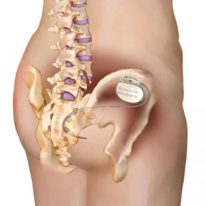
Sacro Neuromodulation – First Stage

The aim is to alter the neurotransmission from the Spinal Centre to the Bladder via the S3 nerve

Why is it done?

  • To alter the neurotransmission from the spinal centre to the bladder:
  • Refractory overactive bladders with urge incontinence (OAB)
  • Underactive bladders (UAB)
  • Chronic pelvic pain
  • Fecal incontinence
  • Causative factors
    • Undetermined
    • Neurogenic causes such as Multiple Sclerosis
  • When at least 2 anticholinergic drugs or B-adrenergic drugs have failed to have provided an improvement in symptoms of OAB
  • An alternative for ISC or permanent IDC for UAB
  • The aim was to alter the neurotransmission from the Spinal Centre to the Bladder
  • This will be a trial to see if this works for you


How is it done?

  • Sedation is administered
  • You will be placed prone (on your stomach) with lower back and buttocks exposed
  • A needle will be placed in the S3 foramina of the sacrum and connected to an electrical current with increased frequency until the correct nerve response is obtained
  • The correct response would be puckering of the anal sphincter as well as the movement of the big toe
  • The lead is then tunneled under the skin
  • The lead is attached to an external modulator and battery.
  • Pts with UAB may have permanent lead placement from the start, as effects may take up to 12 months to occur
  • If you have an OAB: you should experience a marked improvement over the previous 2 weeks
  • A minimal requirement of at least 50% improvement in urinary symptoms is 1required to progress to a full implant
  • As routine in my practice, the permanent lead is used as the temporary, therefore allowing for the exact same results as with the trial period
  • It will be connected to an external battery
  • Generally, a temporary lead is not done for an UAB, as the response may take up to 9-12 months
  • Leads for pain can be placed bilaterally and in multiple sites


Complications

  • Some local discomfort may be experienced.
  • Nerve stimulator may provide abnormal sensations, which your body adjusts to.
  • A representative from Medtronic will be in contact with you to check on your settings and responses.
  • If after a 2-week period of the temporary leads have shown an improved in your bladder, consideration will be given to a permanent implant
  • If no response is obtained the leads may be removed.
  • NB! Each person is unique and for this reason symptoms may vary!


Download Information Sheet


Wes Sacro Neuro Modulation-First Stage Temporary Leads

Copyright 2019 Dr Jo Schoeman

Flexible Cystoscopy

An atraumatic endoscopic procedure to view the bladder. Under local or sedation

A diagnostic day procedure under local anaesthetic, where a flexible cystoscope is placed in the bladder via the urethra.

Why is it done?

To investigate:

  • Haematuria (blood in the urine).
  • Recurrent urinary tract infections.
  • Space occupying lesions in the kidneys, ureters and bladder.
  • Abnormal cells suggestive of urothelial carcinoma, on urine cytology.

Risk factors:

  • Strong family history of bladder cancer.
  • Smokers or passive smokers.
  • Factory workers: dyes, paints, etc.
  • Exposure to Schistosoma (Bilharzia).
  • Renal stone disease, bladder stones.

How is it done?

  • A cystoscopy is performed by placing a camera in the urethra with the help of a lubricant jelly and saline.
  • The bladder is then distended using the fluid.
  • The inside of the bladder is viewed for pathology.
  • If any suspicious lesions are seen, a biopsy will be taken.
  • Urine would have been sent for cytology prior to the procedure, to rule out the existence of cancer.
  • Antibiotics may be given to prevent infection.

What to expect after the procedure?

  • Pain on initial passing of urine.
  • Bladder infection ranging from a burning sensation to, fever, to puss (rare).
  • Blood stained urine.
  • Lower abdominal discomfort which will persist for a few days.
  • NB! Each person is unique and for this reason symptoms vary.

What next?

  • This all depends on what is found during the procedure. All the options will be discussed in detail.
  • With the removal of stents, the ureters have been dilated and will regain function (peristalsis) as soon as the stents are out. Thus slight pain can be expected in the first 24-48hrs.
  • There may be some blood in the urine. This can be remedied by drinking plenty of fluids until it clears.
  • A ward prescription will be issued to patients on discharge, for own collection at any pharmacy.
  • Patients should schedule a follow-up appointment within 7 days.
  • Please don’t hesitate to direct all further queries to Jo.
  • REMEMBER: THOSE WHO SUFFER IN SILENCE, SUFFER ALONE!

Download Information Sheet

Wes Flexible Cystoscopy

Wes Flexible Cystoscopy and Removal Stent

Flexible Cystoscopy & Removal Stent

A day procedure under local anaesthetic, where a flexible cystoscope is placed in the bladder via the urethra to remove a stent placed with previous upper urinary tract work

Why is it done?

To investigate:

  • Removal of stent which was placed after a stone removal, recent ureteroscopy, ureteric re-implantation, precautionary placement prior to pelvic surgery (Colo-rectal, Gynae Oncology, Uro-Oncology)


Risk factors:

  • Strong family history of bladder cancer
  • Smokers or passive smokers
  • Factory workers: dyes, paints, etc
  • Renal stone disease, bladder stones with recent surgery resulting placement of a stent


How is it done?

  • A cystoscopy is performed by placing a camera in the urethra with the help of a lubricant jelly and saline
  • The bladder is then distended using the fluid
  • The inside of the bladder is viewed for pathology.
  • If any suspicious lesions are seen, a biopsy will be taken.
  • Urine would have been sent for cytology prior to the procedure, to rule out the existence of cancer.
  • Antibiotics may be given to prevent infection.
  • Stent removed


Complications

What to expect after the procedure?

  • Pain on initial passing of urine
  • Pain as the ureter contracts back to its usual size
  • Bladder infection ranging from a burning sensation to, fever, to puss (rare)
  • Bloodstained urine
  • Lower abdominal discomfort which will persist for a few days
  • NB! Each person is unique and for this reason, symptoms vary.


Indications for a Ureteric stent

  • Hematuria from upper tracts
  • Disobstruction of the ureter caused either calculus, blood clot or tumour
  • External compression of the ureter by   retro-peritoneal pathology ie: Fibrosis,  retroperitoneal lymph node compression
  • Reduced renal function associated with  hydronephrosis
  • Sepsis associated with hydronephrosis


Download Information Sheet

Wes Flexible Cystoscopy and Removal Stent

Copyright 2019 Dr Jo Schoeman

Flexible Cystoscopy & Urethral Dilation

A day procedure under local anaesthetic, where a flexible cystoscope is placed in the bladder via the urethra. Narrowing in the urethra is dilated.

Why is it done?

A cystoscopy is used to investigate:

  • Hematuria (blood in the urine)
  • Recurrent urinary tract infections
  • Dilatation of Urethral narrowing/ stricture
  • Abnormal cells suggestive of urothelial carcinoma, on urine cytology

Ideally a retrograde urethragram is used to diagnose this radiologically


Risk factors for strictures:

  • Straddle injuries
  • Catheterization or urethral instrumentation
  • Infections
  • Bypass cardiac surgery with long ischemic time


How is it done?

  • A cystoscopy is performed by placing a camera in the urethra with the help of a lubricant jelly and saline
  • Usually, you can’t move past the narrowing
  • Then:

Urethral Dilatation

  • If you have a urethral stricture, a guidewire will be placed and the narrowing dilated
  • There may be some hemorrhaging and you may need a catheter for 3 days
  • This will be removed at the hospital in 3 days or alternatively arrange for your GP to remove.
  • I will review in 6 –8 weeks

Antibiotics may be given to prevent infection

Complications

What to expect after the procedure?

  • You may be sent home with an indwelling catheter for 3 days
  • Pain on initial passing of urine after it is removed
  • Bladder infection ranging from a burning sensation to, fever, to puss (rare)
  • Bloodstained urine
  • Lower abdominal discomfort which will persist for a few days
  • NB! Each person is unique and for this reason, symptoms vary.


Download Information Sheet

Wes Flexible Cystoscopy and Urethral Dilatation IDC

Copyright 2019 Dr. Jo Schoeman

Sacro Neuromodulation – Full Implant

The aim is to alter the neuro-transmission from the Spinal Centre to the Bladder

Why is it done?

  • Confirmed OAB where a trial not required
  • Detrusor Sphincter Dyssynergia, DSD
  • Incomplete bladder emptying, large post-void residual urine volumes
  • Underactive bladders
  • Chronic pelvic pain
  • Causative factors:
    • Undetermined
    • Neurogenic causes such as Multiple Sclerosis,
    • Diabetes

How is it done?

  • Sedation is administered with a local anesthetic of the wound sites
  • You will be placed prone (on your stomach) with lower back and buttocks exposed
  • Area prepared with Betadine, unless allergic
  • The tined lead will be placed in the S3 foramina of the sacrum under imaging
  • The lead will be tunneled under the skin and connected to an Internal Battery
  • A pocket will be made for the permanent subcutaneous battery and connection
  • Subcutaneous sutures will be placed, which will dissolve.


Complications

  • Some local discomfort may be experienced.
  • A nerve stimulator may provide abnormal sensations, which your body adjusts to.
  • A Representative from Medtronic will be in contact with you to check on your settings and responses.
  • A review at my rooms with Medtronics will be scheduled at 6 months, to confirm that the device is functioning optimally
  • Further review appointments will be scheduled as required
  • The battery should last between 15 years
  • NB! Each person is unique and for this reason, symptoms may vary!


Download Information Sheet

Wes Sacro Neuro Modulation Full Implant

Copyright 2019 Dr Jo Schoeman

Hydrocoelectomy

Surgical removal of hydrocele via a scrotal incision with spermatic cord block

Why is it done?

  • Enlarged scrotum.
  • Could be uncomfortable.
  • Usually large and uncomfortable.
  • Can become so big that it buries the penis making usual functions difficult, ie urination and sexual function.
  • May contribute to Infertility.

How is it done?

  • This procedure is done under general anaesthetic.
  • Supine position.
  • A midline scrotal incision is done.
  • The intact hydrocele is delivered through the skin incision.
  • The sac (tunica vaginalis) is opened and surgically removed.
  • A hemostatic running suture is placed around the raw edge of the Tunica Vaginalis.
  • Hemostasis is actively chased.
  • A drain is left overnight.
  • An Indwelling catheter is left for 6-8 hours to prevent acute urinary retention.
  • The scrotum is closed in 2 layers with dissolvable sutures.
  • You would be required to bring 2 pairs of tight new undies for post-operative scrotal support; these will be placed post-operatively.

NB! You are required to bring 2 pairs of tight new undies for post-operative scrotal support.

What to expect after the procedure?

  • Any anesthetic has its risks, and the anesthetist will explain such risks.
  • Bleeding is a possible complication therefore the scrotal drain/s overnight.
  • Your catheter will be removed the next.
  • You will have scrotal swelling and bruising for the next 2-6 weeks.
  • Any sudden increased swelling needs urgent attention!
  • Any symptoms of fever and signs of infection, requires urgent attention!

What next?

  • Dressings should be kept for the initial 72 hours after surgery and soaked off in a bath thereafter.
  • The catheter will be removed the morning after surgery.
  • Patients should schedule a follow-up appointment with Dr Schoeman 4-6 weeks after the procedure.
  • There will be signs of bruising for at least 2-6 weeks.
  • PLEASE CONTACT THE HOSPITAL DIRECTLY WITH ANY POST-OPERATIVE CONCERNS AND RETURN TO THE HOSPITAL IMMEDIATELY SHOULD THERE BE ANY SIGNS OF SEPSIS.

Download Information Sheet

Wes Hydrocelectomy

Indwelling Urethral Catheter – IDC

Non-invasive placement of a silicone tube which is secured inside the bladder and attached to a drainage bag on the outside, in order to drain an obstructed bladder (urinary retention)

Why is it done?

  • This can be placed as an emergency for patients in acute urinary retention
    • Prostate obstruction
    • Urethral strictures
    • Blood clot obstruction caused by bleeding
    • Hematuria (bleeding)
    • Severe urinary tract infections
  • Commonly placed intra-operatively for long, non-urological surgical procedures to enable urine drainage and monitoring urine output.
  • Commonly placed at the end of a Urological procedure to enable urine drainage and to enable hemostasis (stopping bleeding)


How is it done?

  • This is done as a sterile procedure; therefore, the genital area will be cleaned with a non-abrasive disinfectant.
  • A sterile catheter will be used
  • A local anesthetic gel is placed in the urethra a few minutes prior to the placement of the catheter. This may initially sting for a few seconds until it numbs the mucosa.
  • An appropriate size catheter (14-18Fr) will be inserted
  • Urine should be aspirated with a syringe to confirm the correct position in the bladder.
  • An anchoring balloon will be inflated with 10cc of sterile water.
  • A drainage urine bag will be attached
  • The catheter will be secured to your leg. (check that this is always secured)


Complications

  • Urethra with resulting discomfort.
  • In the presence of urethral stricture, it may be impossible to pass the catheter, and a flexible cystoscopy with dilatation of the stricture may be required prior to placement.
  • If you had a large over-stretched bladder (urine retention) you may experience bleeding as the bladder empties, caused by the mucosal tears that have occurred.
  • Catheters that have been placed long term, may cause irritation and possibly attract infection. Permanent catheters are usually changed every 6-8 weeks.


Download Information Sheet

Wes Catheters Indwelling Catheter

Copyright 2019 Dr Jo Schoeman

Intravesical BCG-Therapy

Why is it done?

  • Treatment for localized superficial Urothelial Carcinoma of Bladder and Ureter (T1G3)


How is it done?

  • A Local anaesthetic gel is administered as for a Urethral Catheterization procedure
  • This is done under a sterile procedure.
  • A 14-16 Fr Indwelling Catheter is placed into your bladder.
  • The BCG is installed using strict administering criteria
  • Usually, 1 vial of BCG is mixed with Saline to a 50cc volume
  • The catheter is then removed
  • The BCG is required to stay in your bladder for 2 hours.
  • Body rotation every 30 minutes allows optimal contact of urothelial Surfaces to the BCG.
  • WARNING: Any Fevers require urgent attention


 What next?

  • This will be done every week for 6 weeks
  • 6 weeks after this a check Flexible Cystoscopy will be scheduled as part of your surveillance protocol for your Urothelial carcinoma
  • A Further 2 Installations will be arranged in the following 3 months as part of a    Maintenance Protocol
  • This may be repeated.


Complications

Side–effects

  • Some local discomfort may be experienced.
  • Your voiding nature will change within the next week
  • You may experience some urinary frequency
  • You could develop a fever requiring urgent attention.
  • Systemic effects of BCG would be fever
  • Delayed effects would a urinary tract infection
  • The possibility of Miliary Tuberculosis
  • NB! Each person is unique and for this reason, symptoms may vary!


Download Information Sheet

Wes Intravesical BCG Therapy

Copyright 2019 Dr. Jo Schoeman

Intravesical Botox

Why is it done?

  • To alter the neurotransmission from the nerve to muscle receptor.
  • Refractory overactive bladders with urge incontinence.
  • Chronic pelvic pain?
  • Causative factors:
    • Undetermined
    • Neurogenic causes such as Multiple Sclerosis.
    • Non-neurogenic.
  • NOT INDICATED FOR A PATIENT WITH A HIGH POST VOID RESDUAL URINE VOLUME

How is BOTOX administered

  • A sedation/local anesthetic is administered.
  • You will be placed supine with legs in comfortable frog position
  • A flexible cystoscopy procedure is done.
  • 20 sites of 1 ml blebs are created in a grid fashion with sub-mucosal injections of BOTOX.
  • Usually, 100-200 IU with non-neurogenic bladders and 300 IU with neurogenic causes.
  • Dose may be individualized with subsequent treatments.
  • WARNING: You may not be able to pass urine for up to 2 weeks on your own and may require intermittent self-catheterization. Therefore, if you are not willing to self-catheterize, this is not for you

After the procedure?

  • You will be assessed to see whether you empty your bladder completely.
  • ISC may be instituted if you cannot void or residuals are more than 300cc.

What to expect after the procedure

  • Some local discomfort may be experienced.
  • Your voiding nature will change within the next week to 10 days with a slow stream which may require ISC in < 2% of patients.
  • Systemic effects of BOTOX would cause muscle weakness with higher doses.
  • If you require ISC, this may be required for 2-4 weeks, it will improve thereafter.
  • Neurogenic bladder patients will still continue with ISC as before.
  • NB! Each person is unique and for this reason symptoms may vary!

What next?

  • A date will be set for a review by myself on regular intervals.
  • Please don’t hesitate to direct all further queries to Dr Schoeman’s rooms.


Download Information Sheet

Wes Intravesical BOTOX

Intravesical Mitomycin C –Therapy

Why is it done?

  • Treatment for localized Superficial Urothelial Carcinoma of Bladder and Ureter


How is it done?

  • A Local anesthetic gel is administered as for a urethral catheterization procedure
  • This is done under a sterile procedure.
  • A 14-16 Fr Indwelling Catheter is placed into your bladder.
  • The pre-made-up Mitomycin solution is installed using strict administering criteria
  • Usually, 2 vials of 20mg MMC is mixed with Saline to a 50cc volume. You require 40mg.
  • The catheter is then removed
  • The MMC is required to stay in your bladder for 1 hour.
  • Body rotation is not required.
  • Can also be placed after a bladder tumour resection (TURBT)
  • WARNING: Any Fevers require urgent attention


 What next?

  • Some local discomfort may be experienced.
  • Your voiding nature will change within the next week
  • You may experience some urinary frequency
  • You could develop a fever requiring urgent attention.
  • Some patients may experience severe pain when/ if the tumour was resected very deep.

NB! Each person is unique and for this reason, symptoms may vary


Complications

Side–effects

  • This will be done every week for 6 weeks
  • 6 weeks after this a check Flexible Cystoscopy will be scheduled as part of your surveillance protocol for your Urothelial carcinoma
  • This may be repeated.
Surveillance Protocol for Superficial Urothelial Carcinoma

Low Grade: Ta, T1 (G1, G2), CIS

· Initially 3 months after the first resection

· If clear then 9months

· Then annually

High-Risk Low Grade: T1G3 with/out CIS, high volume disease

· This disease may be best treated with a radical cystectomy

· 3 monthly check cystoscopy for 12 months

· If clear, then 6 monthly for a further 12 months

· If clear then annually for 5-7 years

· My recommendation thereafter would be a 2-yearly cystoscopy with intermittent Urine Cytology

· You fall back to the beginning with any recurrences


Download Information Sheet

Wes Intravesical MMC Therapy

Copyright 2019 Dr. Jo Schoeman