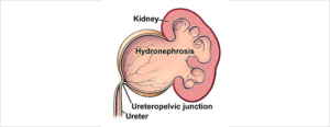
Pelvi-Ureteric Junction Repair (PUJ) – Robotic Assisted

A congenital or acquired narrowing in the ureteric pelvis junction. This narrowing is excised with a reconnection. There are several techniques described in repairing this: I prefer the Dismembered Pyeloplasty

Why is it done?

  • High grade obstruction.
  • Causing deterioration of renal function.
  • Thinning of renal cortex.
  • Chronic pain.
  • Chronic infection.
  • Recurrent renal calculi.

Causes

  • Congenital lack of muscle, or neuro transmission in this area, causing a non-functioning part leading to obstruction.
  • Vesico-ureteric reflux, longstanding can also cause this.
  • Usually diagnosed in kids.
  • Crossing vessel.

How is it done?

Robotic assisted pyeloplasty.

  • Types
      • Dismembered.
      • Foley’s Y-V Pyeloplasty.
      • Culp-Dewierd.
      • Pelvi-calyceal pyeloplasty.
      • Endopyelotomy with laser.
    • Patients will receive a general anaesthesia.
    • Prophylactic antibiotics is given.
    • The correct ureteric system is identified and marked while you are awake.
    • This will be mostly a robotic / laparoscopic procedure.
    • The endoscopic procedure is reserved as a second line in my practice.
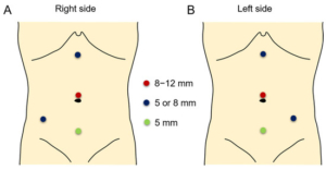
    • Laparoscopic ports are placed
    • The affected ureter is exposed, the defect cut out with a re-anastomosis of a spatulated ureter to a trimmed renal pelvis over a ureteric stent.
    • An indwelling catheter is placed. A drain is placed.

What next?

  • You may be in hospital for 3 days
  • Your drain will be removed when there is no urine draining.
  • Your catheter will be removed the following day.
  • As soon as you are comfortable with no signs of pain and emptying your bladder sufficiently, you will be discharged
  • A ward prescription may be issued on your discharge, for your own collection at any pharmacy.
  • A follow-up appointment will be scheduled for 6 weeks to remove your ureteric stent under local anaesthesia with a Flexible Cystoscope.
  • A review with a CT IVP will be scheduled 6 weeks after this to check on the end result of the ureter.
  • Any pain or signs of fever require an urgent review.
  • DON’T SUFFER IN SILENCE, OR YOU WILL SUFFER ALONE!

Possible Complications

  • Re-stenosis with recurrent obstruction.
  • Second procedure.
  • With further deterioration of renal function, you may require a nephrectomy where affected kidney contributes < 15-20% of total renal function.
  • Urine leak, Urinoma, requiring drainage.
  • Infection possible sepsis requiring long-term antibiotics.


Download Information Sheet

Wes RA reair

Laser Endo-Pyelotomy

Endoscopic opening of pelvi-ureteric junction stricture.

Endoscopic technique of incising a short stricture with a laser. A stent remains 6 weeks post operatively

Why is it done?

  • Stricturing or narrowing of the ureter causing significant hydronephrosis.
  • Leading to chronic infection.
  • End-result is loss of renal function.
  • An end-to-end anastomosis can be considered in the mid ureter.
  • A Reimplantation into the bladder with lower ureteric strictures.
  • A pelvi-ureteric junction repair in higher ureteric strictures.
  • A trans uretero-ureteric anastomosis joining one ureter to the other where long defects are present.
  • Renal Auto Transplantation where ureter is completely damaged or.
  • An ileal ureteric substitution where to whole ureter is damaged.

How is it done?

  • Endoscopic technique.
  • Patients will receive a general anesthesia.
  • Prophylactic antibiotics is given.
  • The correct ureteric system is identified and marked while you are awake.
  • This will be mostly an endoscopic procedure.
  • A cystoscopy will be done with placement of ureteric guidewires.
  • The affected ureter is entered with ureteroscopy. Laser is used to cut through the short stricture until peri-ureteric fat is observed.
  • This is not an advisable procedure where the stricture overlies vascular structures.
  • A ureteric stent is placed.
  • An indwelling catheter is placed.

What next?

  • You may be in hospital for 3 days.
  • Your drain will be removed when there is no urine draining.
  • Your catheter will be removed the following day.
  • As soon as you are comfortable with no signs of pain and emptying your bladder sufficiently, you will be discharged.
  • A follow-up appointment will be scheduled for 6 weeks to remove your ureteric stent under local anesthesia with a Flexible Cystoscope.
  • A review with a CT IVP will be scheduled 6 weeks after this to check on the end result of the ureter.
  • Any pain or signs of fever require an urgent review.
  • DON’T SUFFER IN SILENCE, OR YOU WILL SUFFER ALONE!

Download Information Sheet

Wes Laser Endo-Pyelotomy

Greenlight Laser Trans Urethral Vaporization of Prostate

This is an endoscopic technique of enucleation of the prostate using Greenlight laser therapy. Similar to a TURP – “Re-bore” only with minimal bleeding and shorted hospital stays. Some would argue this is ” The new Gold Standard”

Why is it done?

Endoscopic vaporization of a benign enlarged prostate, using laser.

Indications:

  • Patients on anticoagulation / anti-platelet therapy
    • Warfarin needs to be placed on Clexane 7 days prior.
    • Clopidogrel should be down scaled down to Aspirin.
    • This allows patients on anti-coagulation therapy to continue their medication with minimal risk of hemorrhage. It also allows a shorter stay in hospital.
  • Prostates up to 120cc.
  • Where conservative management has failed.
  • Patient choice.
  • This procedure is performed when the prostate gland is enlarged to such an extent that medication cannot relieve the urinary symptoms.
  • Symptoms include: LUTS
    • a weak stream,
    • nightly urination,
    • frequent urination,
    • inability to urinate,
    • kidney failure due to the weak urination (obstruction),
    • bladder stones,
    • recurrent bladder infections.
  • Medication such as Flomaxtra, Urorec or Minipress etc. should always be given as a first resort.
  • Step-up therapy could have been used for prostates larger than 35-50cc where indicated, with either Duodart, Avodart or Proscar.
  • Prostate cancer first needs to be ruled out by doing a PSA, and when indicated, with a 3T MRI scan of the prostate with an abnormal PSA with a possible prostate biopsy of any suspicious lesions.

How is it done?

    • Patients will receive a general anesthesia, unless contra-indicated.
    • A cystoscopy is performed by placing a camera in the urethra with the help of lignocaine gel.
    • Saline is used to irrigate the bladder, therefore NO DILUTIONAL HYPONATRAEMIA
    • The inside of the bladder is viewed for pathology. If any suspicious lesions are seen, a biopsy will be taken.
    • A vaporization of the prostate is then started and should take 60-120 minutes depending on the size of the prostate.
    • Prophylactic antibiotics will be given to prevent any infections.
    • Post– operative antibiotics will be continued for 10 days.
    • No specimen will be obtained due to vaporization, unless PSA was suspicious and an MRI with view to prostate biopsy has excluded a prostate cancer.

What can go wrong?

  • Any anesthesia has its risks, and the anesthetist will explain this to you.
  • No blood loss is expected.
  • In rare circumstances you may develop fluid overload requiring a High Care Facility admission, especially with the upper edge of prostate sizes.
  • You will wake up with a catheter in your urethra and bladder. This will remain in the bladder overnight.
  • Lower abdominal discomfort for a few days.
  • NB! Each person is unique and for this reason symptoms vary!

What next?

  • You will spend 1 night in hospital.
  • You will a trial without catheter the next day.
  • You will be discharged as soon as you can completely empty your bladder.
  • You may initially suffer from urge incontinence and dysuria (discomfort) and will improve within the next 6 weeks.
  • You will pass a grey, wet scab at the 2–3-week mark.
  • Some bleeding may be associated with this, especially if you are on anticoagulation therapy.
  • Allow for 6 weeks for stabilization of symptoms.
  • There may be some blood in your urine. You can remedy this by drinking plenty of fluids until it clears.
  • A follow-up appointment will be scheduled for 6 weeks. Remember there is no pathology due to vaporization.
  • DON’T SUFFER IN SILENCE, OR YOU WILL SUFFER ALONE!

Side–effects

  • Retrograde ejaculation in more than 90% of patients. Therefore, if you have not completed your family, this procedure is not for you unless absolutely necessary.
  • Infertility as a result of the retrograde ejaculation. Not a given, therefore continue your contraceptives
  • Stress incontinence especially in the elderly and the diabetic patients.
  • Patients with Multiple Sclerosis, Strokes and Parkinsons have a higher risk of incontinence and risks should be discussed and accepted prior to surgery.
  • Urethral stricturing in <2% of patients, requiring intermittent self-dilatation.
  • Regrowth of prostate lobes can occur within 3-5 years requiring a second procedure.
  • NB! Each person is unique and for this reason symptoms vary!

Remember

  • You still have a peripheral zone of your prostate and regular PSA reviews are required up to the age of 75. (This could be seen as controversial).

Download Information Sheet

Wes TUVP Greenlight

Low Dose Brachytherapy

Perineal placement of Radioactive Iodine-125 seeds in the prostate as treatment for Intermediate-Risk Prostate Cancer

Why is it done?

This is a radiation therapy option for the management of a localized prostate cancer.

Criteria include:

  • PSA less than 10
  • Gleason 3,4 adenocarcinoma prostate,
  • Higher grades may have extra-prostatic extension requiring combined External Beam Radiation
  • Staging negative, (bone scan negative, CT negative)
  • Not younger than 65
  • It is the localized radiation of the prostate, by means of trans-perineal placement of Radio-active I-125 seeds.
  • This is a nerve sparing procedure and patients have a good opportunity to maintain this.
  • The procedure takes 2-3hours excluding the anesthetic time.
  • This is an alternative to a Radical Retropubic Prostatectomy for low-intermediate risk prostate cancer. Prostates are generally smaller than 50cc.
  • Could be ideal for those patients with excessive BMI and fitting the cancer specific criteria.


How is it done?

  • Under a General Anaesthetic
  • Presence of Radiation Oncologist and Physicist
  • Lithotomy position
  • Trans-rectal placement of an ultrasound probe
  • Real-time accumulation of digital images to allow real-time placement of Radioactive Iodine seeds according to an intra-operative plan

PSA failure:

  • PSA not dropping to a nadir value, preferably 0,2ng/ml.
  • 3 consecutive PSA rises following RRP.

Complications

  • Perineal hematoma
  • Wound infections
  • Urgency frequency and weak stream
  • Limited Erectile Dysfunction, which may only surface after 18 months after treatment.
  • Lower ejaculate volume.
  • Testicular pain similar to vasectomy for 2-3 days


Post operative care:

  • A post-procedure CT scan to account all the seeds and exclude migration of seeds.
  • Normal diet
  • A salt water or Betadine Douche is required after every stool for the first week
  • A 3-month course of Flomaxtra to ease Lower Urinary Tract Symptoms
  • Wounds generally heal in 7-10 days

Catheter care

  • Your catheter will remain until you are awake.
  • Post-operative review:
  • Radio-Oncology will review a few weeks later.
  • You will review with me at 6 weeks post-procedure to check if you are doing fine.
  • Review PSA roughly 3 months after the procedure to assess PSA track
  • You should reach your Nadir (lowest PSA) at 6– 9 months after the procedure. The lower the better.
  • 6 monthly PSAs thereafter.
  • Expect a PSA Bump at approximately 9-12 months after the procedure, The PSA should drop thereafter
  • If stable with good PSA outcomes, refer back to GP for 6 monthly PSA review


Download Information Sheet

Wes Low Dose Brachytherapy

Peri-Urethral Bulking Injections

Periurethral bulking for a type 3 urinary incontinence: “lead-pipe urethra” (ISD): Bulkamid or Macroplastique


Why is it done?

  • Where Intrinsic Sphincter Deficiency has been proved after failed previous sling.

How is it done?

  • This procedure is done under a spinal / general anesthetic, as decided by the anesthetist.
  • The legs will be elevated into the lithotomy position.
  • This procedure is done cystoscopically.
  • 3 peri-urethral injections are made with injection of Macroplastique/ Bulkamid until the urethral lumen closes.
  • Your urine output will be measured each time you urinate, and the residual will be measured. (Patients will be required to do this up to 3 times.)
  • If the residual amount of urine is more than 1/3 of the total bladder capacity, the patient may have to self-catheterize, until the residual volume is acceptable.
  • Prophylactic antibiotics will be given to prevent infection.

What to expect after the procedure?

  • Any anesthetic has its risks, and the anesthetist will explain all such risks.
  • Complications of hemorrhaging and urine retention.
  • Patients will have no catheter when they wake up.
  • If you cannot urinate after 2-3 attempts, an in-out catheter may be inserted to empty your bladder.
  • You may be required to self-catheterize for a week or two.
  • NB! Each person is unique and for this reason symptoms may vary!

What next?

  • Patients will have a trial of void without catheter after the procedure
  • Patients will be discharged as soon as they can completely empty the bladder.
  • Patients may be required to self-catheterize for a week or two (rarely necessary).
  • Patients may initially suffer from urge incontinence, but this will improve within the next 6 weeks.
  • Allow 6 weeks for symptoms to stabilize.
  • You may not experience a full return of continence, and the effects may worsen with time.
  • More than 1-2 treatments may provide the desired effects.
  • There may be some blood in the urine. This can be remedied by drinking plenty of fluids until it clears.
  • On discharge a prescription may be issued for patients to collect.
  • Patients are to schedule a follow-up appointment in 6 weeks.
  • Please direct all queries to Dr Schoeman’s rooms.
  • PLEASE CONTACT THE HOPSITAL DIRECT WITH ANY POST-OPERATIVE CONCERNS AND RETURN TO THE HOSPITAL IMMEDIATELY SHOULD THERE BE ANY SIGNS OF SEPSIS.


Download Information Sheet

Wes Peri-Urethral Bulking

Male Advance Sling

For those guys who suffer persistent low-intermediate grade of stress urinary incontinence after their radical prostatectomy for prostate cancer. Usually 2 level 2 pads per day.

Why is it done?

  • Male Stress incontinence
  • Usually after a TURP/TUVP, Radical Prostatectomy in 2% of cases.


How is it done?

  • This procedure is done under a spinal / general anesthetic, as decided by the anesthetist.
  • The legs will be elevated into the lithotomy position.
  • A 5-7cm incision is made on the perineum (space between scrotum and anus).
  • The sling is placed around the bulb of the penis.
  • The arms of the sling are brought to the skin at the inner thigh, with a small incision.
  • The sling is placed with descent tension, pulling the bulb inwards by at least 2 cm.
  • The bladder will be inspected with a cystoscopy to exclude any injuries to the bladder wall and urethra.
  • The wounds are closed with dissolvable sutures.
  • A local anesthetic is given for pain relief.
  • A urinary catheter is placed for 24hrs.
  • The catheter will be removed early the next morning.
  • The patient’s post void residual volume will be measured. (Patients will be required to do this up to 3 times.)
  • If the residual amount of urine is more 250-300 cc the patient may have to self-catheterize, until the residual volume is acceptable.
  • You may require an indwelling catheter for 7 days.
  • Prophylactic antibiotics will be given to prevent infection.


What to expect after the procedure?

  • Any anesthetic has its risks, and the anesthetist will explain all such risks.
  • Complications: hemorrhaging, requiring blood transfusion <1%.
  • Patients will wake up with a catheter in the urethra and bladder. This will remain in the bladder for 24 hrs.
  • Inner thigh discomfort/pain will persist for a few days but this will subside / settle.
  • If you cannot urinate after 2-3 attempts, the catheter may be replaced for a further 7 days
  • You may be required to self catheterize for a week or two.
  • If there is no improvement the sling may be cut, to allow spontaneous urination
  • This may only be 50% effective in irradiated patients
  • NB! Each person is unique and for this reason symptoms may vary!


Download Information Sheet

Wes Male Advance Sling

Meatotomy

Opening of a meatal stenosis (pinhole narrowing).

Why is it done?

  • To treat a narrowing in the tip of the urethra which has formed due to previous damage/injury to the urethra.
  • Where intermittent dilatation is not desired, as discussed with the patient.
  • Causes:
    • Circumcision in early childhood where the foreskin is still attached to the glans, usually neonatal circumcision
    • After bypass surgery where a drop in blood pressure has caused an area of low blood supply to the urethra.
    • trauma to the urethra (pelvic fractures/ urethral instrumentation).
    • and sexually transmitted diseases.
  • The procedure involves surgical refashioning of your meatus.

How is it done?

  • Usually done in male patients
  • Selected Female patients can be considered.
  • Patients will receive a general anesthetic.
  • Your meatus will be refashioned, in order to leave it patent without a narrowing.
  • An indwelling catheter is left till post-operative period.
  • Prophylactic antibiotics may be given to prevent infection.

What to expect after the procedure?

  • It may be slightly uncomfortable.
  • A catheter will be inserted in the urethra and bladder. This will remain in until you are awake.
  • Catheters can be very irritating and cause some discomfort.
  • Blood stained urine will be present.
  • Painful urination may persist for a few days.
  • NB! Each person is unique and for this reason symptoms may vary!

What next?

  • Patients will be sent home after a successful attempt at voiding.
  • You may experience some discomfort with every void, that will become less over the next few voids.
  • There may be some blood in the urine. This can be remedied by drinking plenty of fluids until it clears.
  • On discharge a prescription may be issued for patients to collect.
  • Patients should schedule a follow-up appointment with Dr Schoeman within 6-8 weeks.
  • Should patients have any problems with urination, please contact the rooms for an earlier appointment.
  • Please don’t hesitate to direct any further queries to Dr Schoeman’s rooms.
  • PLEASE CONTACT THE HOSPITAL WITH ANY POST-OPERATIVE CONCERNS AND RETURN TO THE HOSPITAL IMMEDIATELY SHOULD THERE BE ANY SIGNS OF SEPSIS.

Download Information Sheet

Wes Meatotomy

Trans Urethral Resection Prostate (TURP) – Monopolar

This is the procedure used to resect the inside (enlarged, obstructive part) of the prostate. Known generally as the “Re-Bore”.  Glycine is used as an irrigate.                                                           

Why is it done?

  • This procedure is performed when the prostate gland is enlarged to such an extent that medication cannot relieve the urinary symptoms.
  • Symptoms include
    • a weak stream,
    • nightly urination,
    • frequent urination,
    • inability to urinate,
    • kidney failure due to the obstruction,
    • bladder stones,
    • recurrent bladder infections due to retaining urine.
  • Medication such as Flomaxtra, Urorec or Minipress etc. should always be given as a first resort.
  • Step-up therapy could have been used for prostates larger than 35-50cc with either Duodart, Avodart or Proscar, where indicated and appropriate.
  • Prostate cancer first needs to be ruled out by doing a PSA, and when indicated, with a 3T MRI scan of the prostate with an abnormal PSA with a possible prostate biopsy of any suspicious lesions.

How is it done?

  • Patients will receive a general anesthesia, unless contra-indicated.
  • A cystoscopy is performed by placing a camera in the urethra with the help of a lubricant jelly and an irrigant fluid.
  • The inside of the bladder is viewed for pathology. If any suspicious lesions are seen, a biopsy will be taken.
  • A resection of the prostate is then started and should take 60-90 minutes.
  • Prophylactic antibiotics will be given to prevent any infections.


Complications

Side–effects

  • Retrograde ejaculation in more than 90% of patients. Therefore, if you have not completed your family, this procedure is not for you unless absolutely necessary.
  • Infertility as a result of the retrograde ejaculation.
  • Stress incontinence especially in the elderly and diabetic patients
  • Patients with Multiple Sclerosis, Strokes and Parkinsons have a higher risk of incontinence and risks should be discussed and accepted prior to surgery.
  • Urethral structuring in 5% of patients, requiring intermittent self-dilatation.
  • Regrowth of prostate lobes within 3-5 years requiring a second procedure.

NB! Each person is unique and for this reason, symptoms vary.


Download Information Sheet

Wes TURP Monopolar

Copyright 2019 Dr Jo Schoeman

Focal Therapy for Prostate Cancer – NanoKnife

Not covered by Medicare / Health Fund yet, possibly early 2026

What is IRE

  • Irreversible Electroporation Therapy—breaking up of cell membranes using electric current by means of creating holes in the cell walls (Nano-pores)
  • Non-thermal ablation


Why is it done?

  • Treatment for localized prostate cancer
  • Single focus disease
  • Prostate preserving
  • Ejaculate sparing
  • Continence preserving


How is it done?

  • Focal therapy of prostate cancer, usually a single lesion
  • Preferably Gleason 3,4 or more aggressive
  • Preserving prostate supporting tissue and erectile function and continence

You would have had a 3T MRI study possibly a PET PSMA as well prior to confirming your prostate cancer. Usually, whole gland biopsies are taken of the prostate via the perineum to prove unifocal cancer.

  • This procedure is done under general anesthesia as a day procedure and takes approximately 60-90min (incl anesthetic time)
  • It is performed with the patient lying in lithotomy (legs in stirrups) position
  • Prophylactic antibiotics are essential and a script with details is provided on the day of signing consent.
  • A trans-rectal ultrasound is placed
  • 4-5 NanoKnife electrodes are paced approximately 2cm apart surrounding the focal cancer.
  • An electrical current of 3A is run at 800-1331mcs pulse causing a non-thermal ablative technique
  • Total treatment as soon as electrodes are placed is < 5 minutes


Complications

Side–effects

  • Hematuria (blood in urine) 2-3days
  • Hematospermia (blood in ejaculate) will become less the more often you ejaculate.
  • Bacteremia (infection) with low-grade fever and feeling unwell
  • Perineal hematoma
  • Perineal pain and penis tip pain
  • Prostate swelling causing bladder outlet obstruction requiring a catheter for up to 5 days

ANY FEVER REQUIRES URGENT ATTENTION

What next?

  • You will be discharged with an indwelling catheter for 3-5 days depending on the size of your prostate and the lesion treated
  • You may have necrotic tissue developing requiring a resection (treatment involving the urethra).
  • Difficulty in urination up to 6 weeks after the procedure
  • It could feel like you are sitting on a golf ball for a week


Should there be any signs of fever or cold shivers, you are to return to the hospital or Emergency Department without hesitation

Download Information Sheet


Wes Nanoknife

Copyright 2019 Dr Jo Schoeman

One Stage Urethraplasty

Primary excision and anastomosis of a short segment urethral stricture.

Why is it done?

  • To treat urethral strictures (narrowing) caused by trauma, infection, malignancy, etc. Shorter strictures less than 2 cm in length.

How is it done?

  • This procedure is done under general anesthetic.
  • Legs are placed in a lithotomy position.
  • A single incision is made on the midline raphe on the perineum (area between scrotum and anus).
  • The corpus spongiosum identified and exposed
  • Stricture located using urethral sounds
  • The stricture is excised with a spatulated anastomosis over an indwelling catheter
  • A long-term catheter will be inserted for 10 days.
  • A dressing is then applied, which should be removed after 72 hours.
  • A local anaesthetic is injected into the wound, thus giving post-operative pain relief for the next 4-6 hours.
  • A drain may also be left for 24-48 hours to prevent the collection of serous fluids.

What to expect after the procedure?

  • Any anaesthetic has its risks and the anaesthetist will explain all such risks.
  • You will be sent home with an Indwelling catheter for 7-10 days
  • Bleeding is a common complication.
  • A haematoma (blood collection under the skin) may form and needs to be reviewed by Dr Schoeman as soon as possible. Bruising is normal.
  • An infection of the wound may occur and requires immediate attention.
  • Erectile dysfunction (15%) may occur.
  • Re-stricturing (20-30%) may occur.
  • Owing to the area of the surgery the wound should be kept clean and dry.
  • DANGER SIGNS: A scrotum that swells immediately to the size of a football, fever, or puss. Please contact Dr Schoeman or the hospital immediately as this may occur in up to 15 % of all cases.

What next?

  • The dressing should be kept dry for the initial 72 hours after surgery and then soaked in a bath until it comes off easily.
  • The dressing may sometimes adhere to the wound causing slight bleeding on removal. Don’t panic, the bleeding will stop.
  • Arrangements will be made for the removal of the catheter after 7-10 days.
  • A urinating Urethragram will be arranged with radiology within 6 weeks to determine the final result of the surgery.
  • There will be signs of bruising for at least 10 days.
  • The suture-line will be hard and indurated for at least 8-10 weeks.
  • PLEASE CONTACT THE HOSPITAL DIRECT WITH ANY POST-OPERATIVE CONCERNS AND RETURN TO THE HOSPITAL IMMEDIATELY SHOULD THERE BE ANY SIGNS OF SEPSIS.

Download Information Sheet

Wes Urethraplasty One-Stage