Ureteroceles: symptoms, investigations and treatment

A ureterocoele (also spelt ureterocele) is a balloon-like swelling at the lower end of a ureter: the tube carrying urine from the kidney to the bladder. The swelling usually projects into the bladder. Its opening may be narrow, slowing urine drainage and sometimes causing infection, stones or pressure on the kidney.

Some ureterocoeles cause no problems and are found incidentally. Others need treatment to improve drainage and protect kidney function. [1,2]

Congenital or acquired?

Most true ureteroceles are congenital, meaning they develop before birth. They may be detected on pregnancy ultrasound, during childhood or much later in adult life. A diagnosis in adulthood does not necessarily mean the ureterocele developed recently.

A ureterocoele can occur with a single ureter or a duplex collecting system, in which a kidney has two drainage systems. In a duplex kidney, the ureterocoele commonly involves the ureter draining the upper part of the kidney.

An intravesical or orthotopic ureterocoele lies within the bladder. An ectopic ureterocoele extends into the bladder neck or urethra and may interfere with bladder emptying. Adults more often have an intravesical ureterocoele associated with a single drainage system. [1,3]

The term “acquired ureterocoele” needs care. An acquired swelling of the lower ureter that resembles a ureterocoele is often called a pseudoureterocoele. A stone or a lesion involving the bladder near the ureteric opening can cause this appearance. An irregular or unusual swelling should therefore be investigated rather than assumed to be a harmless congenital abnormality. [4]

Presentation and symptoms

A ureterocoele may be discovered during investigations for an unrelated problem. When symptoms occur, they can include:

  • Recurrent urinary tract infections.
  • Pain in the side, back or lower abdomen.
  • Burning, urgency or frequent urination, particularly with infection.
  • Blood in the urine.
  • Stones trapped within the ureterocoele.
  • Difficulty emptying the bladder or, occasionally, urinary retention.

Children may present with unexplained fever, feeding difficulties or poor growth associated with infection. Rarely, a large ureterocoele can prolapse through the urethra.

Symptoms depend on whether the swelling obstructs drainage, contains a stone or affects the bladder outlet. Prolonged obstruction and repeated kidney infections can damage kidney function. [2,5]

What investigations are needed?

Investigations establish the anatomy, assess drainage and kidney function, and look for infection or reflux. Not everyone needs every test.

Investigation What it helps assess
Urine testing and culture Infection and the appropriate antibiotic
Blood tests Kidney function; inflammatory markers when infection is suspected
Kidney and bladder ultrasound The ureterocoele, stones and swelling of the kidney or ureter
CT, sometimes CT urography Stones and detailed urinary tract anatomy, particularly in adults
MR urography Complex anatomy, including duplicated drainage systems
MAG3 or DMSA kidney scan Drainage and relative function, or functioning kidney tissue and scarring
Micturating cystourethrogram (MCUG/VCUG) Urine flowing backwards from the bladder into a ureter
Cystoscopy Direct inspection of the bladder and ureteric openings

CT involves radiation; contrast use depends on kidney function and the clinical question. Reflux testing is particularly relevant in children or when infection, complex anatomy or postoperative concerns warrant it. Cystoscopy can clarify an uncertain diagnosis and allow treatment during the same procedure. [1,3,5]

Does every ureterocoele need surgery?

No. Observation with planned follow-up may be appropriate when there are no troublesome symptoms, significant obstruction or recurrent infections, and kidney function is satisfactory.

Monitoring may include ultrasound and review of symptoms. Antibiotics treat infection but do not correct the swelling. Preventive antibiotics are considered selectively, particularly in children at increased infection risk. [6]

An infected, obstructed kidney requires urgent assessment and drainage, alongside antibiotics. Drainage may involve endoscopic opening of the ureterocoele or a nephrostomy, a tube placed through the skin into the kidney. Antibiotics alone may be insufficient when infected urine cannot drain. [1]

Endoscopic incision or deroofing

Treatment can often be performed through a telescope passed along the urethra into the bladder, usually under general anaesthesia.

Incision or puncture creates an opening in the wall of the ureterocoele. Deroofing removes part of its wall to create a broader opening. Both aim to let urine drain freely and reduce the balloon-like swelling.

The choice depends on anatomy, obstruction and associated stones. Instruments using electrical energy or a laser may be used. A stone within the sac can sometimes be fragmented and removed at the same operation.

A bladder catheter may be needed temporarily. Endoscopic treatment can be sufficient for a straightforward adult intravesical ureterocoele, while complex duplex or ectopic cases may need further surgery. [5,7,8]

Where does a ureteric stent fit in?

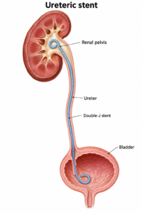
A double-J stent is a soft internal tube extending from the kidney to the bladder. It may be used selectively to support drainage after treatment, during associated stone surgery or while swelling settles. It is not required in every case and does not necessarily provide a permanent solution to the ureterocoele. [8]

Stents can cause frequency, urgency, bladder discomfort, blood in the urine and flank pain, sometimes after urination. Infection, displacement and mineral encrustation are other possible problems.

Every temporary stent needs a clear removal or exchange plan. The duration depends on the procedure and clinical circumstances. Contact the treating team if the removal date is unclear or overdue. [9]

Possible complications, including vesicoureteric reflux

Possible complications of endoscopic treatment include bleeding, infection, discomfort, injury to nearby urinary tract structures, persistent obstruction or narrowing as the opening heals. Repeat treatment or reconstructive surgery may be needed. [3,5,7]

Vesicoureteric reflux (VUR) means urine travels backwards from the bladder towards the kidney. It may already be present before treatment, particularly in a duplex system, or develop after opening the ureterocoele alters the natural anti-reflux mechanism.

Reflux does not automatically mean kidney damage or another operation. Its significance depends on its severity, infections, kidney function and bladder function. Monitoring may be appropriate; persistent significant reflux with recurrent infections can require further treatment, including ureteric reimplantation.

Risk varies with anatomy and technique. Ectopic ureterocoeles and duplex systems have higher rates of postoperative reflux and additional surgery than simpler intravesical cases. [1,7]

In one small adult study, reflux was detected in two of 23 patients followed after endoscopic incision, persisting in one at six months. These results illustrate the possibility of reflux; they do not predict an individual patient’s risk. Adult evidence largely comes from small studies. [7]

Further surgery and follow-up

Reconstructive options include reimplanting the ureter into the bladder or joining it to another healthy ureter in a duplex system. Occasionally, a severely damaged, poorly functioning kidney segment causing problems may require removal. Specialist assessment determines whether reconstruction, observation or removal is appropriate. [1]

Follow-up checks that drainage has improved and looks for recurrent infection, obstruction or reflux. Ultrasound is commonly used; further imaging is selected according to the findings. Symptom improvement alone does not confirm that drainage is normal. [5,7]

Seek urgent medical care for fever or shaking chills with flank pain, inability to pass urine, severe worsening pain, persistent vomiting or feeling seriously unwell. These symptoms may indicate infection or obstruction, including after treatment or with a stent in place. [1,9]

This article provides general education. Treatment should be tailored to the patient’s anatomy, symptoms and kidney function in consultation with their urologist.

References

  1. European Association of Urology. EAU Guidelines on Paediatric Urology: Ureterocele and ectopic ureter. Current online guideline, accessed 5 October 2026. https://uroweb.org/guidelines/paediatric-urology/chapter/ureterocele-and-ectopic-ureter
  2. European Association of Urology. Patient information: Ureterocele. https://patients.uroweb.org/condition/duplex-congenital-malformations-of-the-urinary-tract/ureterocele
  3. The Outcome of Transurethral Electro-Endoscopic Resection of Orthotopic Single System Ureterocoeles in Adults Nigerians. https://pmc.ncbi.nlm.nih.gov/articles/PMC11073836/
  4. Thornbury JR, Silver TM, Vinson RK. Ureteroceles vs. pseudoureteroceles in adults. Urographic diagnosis. Radiology. 1977;122(1):81–84. doi:10.1148/122.1.81. https://pubmed.ncbi.nlm.nih.gov/830358/
  5. An adult ureterocele complicated by a large stone: A case report. International Journal of Surgery Case Reports. 2018. https://pmc.ncbi.nlm.nih.gov/articles/PMC5927814/
  6. European Association of Urology. Watchful waiting for ureterocele. https://patients.uroweb.org/condition/duplex-congenital-malformations-of-the-urinary-tract/ureterocele/wait-and-see-ureterocele
  7. Vijay MK, et al. The safety and efficacy of endoscopic incision of orthotopic ureterocele in adult. Saudi Journal of Kidney Diseases and Transplantation. 2011;22(6):1169–1174. https://pubmed.ncbi.nlm.nih.gov/22089776/
  8. A Large Stone Within a Ureteroceles: A Diagnostic Pitfall and the Utility of Holmium Laser Deroofing as a Viable Surgical Option. https://pubmed.ncbi.nlm.nih.gov/29468199/
  9. British Association of Urological Surgeons. Information about your ureteric stent. https://www.baus.org.uk/_userfiles/pages/files/Patients/Leaflets/Stent%20advice.pdf

Vaginal vault prolapse: understanding the options

Patient information | Reviewed 28 September 2026

Vaginal vault prolapse occurs when the top of the vagina descends after a hysterectomy. It can occur alone or alongside a bulge of the front vaginal wall (anterior prolapse, often called a cystocele). Some women feel a bulge, dragging or pressure; others have difficulty emptying their bladder, recurrent urinary infections, bowel symptoms or discomfort during sex. The size of a prolapse on examination does not always match how troublesome it feels. Treatment should be guided by symptoms and personal priorities, rather than the examination alone. [1,2]

Why the front wall matters

The vaginal apex helps support the front and back walls. An anterior bulge may partly reflect loss of support at the apex. Repairing only the front wall while leaving significant vault prolapse untreated may leave the underlying problem unresolved. Conversely, restoring apical support may improve some anterior prolapse, although a separate anterior repair is sometimes needed. The surgeon should assess each compartment before deciding which parts require treatment. [1,3]

Assessment and treatment without surgery

Assessment includes a history of bulge, urinary and bowel symptoms, sexual function, prior hysterectomy and prolapse operations; examination while straining; and discussion of what the woman wants treatment to achieve. A bladder scan for residual urine is useful when emptying is difficult. Urine tests, urodynamics or imaging are considered for specific questions rather than required for every woman. Stress leakage can be hidden by a large prolapse and may appear after it is reduced or repaired; this possibility deserves discussion before surgery. [1,2]

If symptoms are mild, observation is reasonable. Pelvic floor muscle training can improve symptoms, particularly with less advanced prolapse, but cannot reliably lift a substantial vault prolapse back into place. A vaginal pessary can relieve a bulge without surgery and can also help someone decide whether restoring support improves bladder or bowel symptoms. Pessaries require fitting and ongoing review. Vaginal oestrogen may help postmenopausal vaginal dryness or irritation when appropriate; it is not a cure for the prolapse. [1,2]

Surgical options

Approach How the apex is supported Main considerations
Vaginal sacrospinous fixation Stitches attach the vaginal top to a pelvic ligament, usually on one side. Avoids an abdominal incision and implanted mesh; possible temporary buttock pain and recurrent anterior bulge.
Vaginal uterosacral ligament suspension Stitches attach the vaginal top to the uterosacral ligaments. Uses the woman’s own tissues; attention to the ureters is essential, often with cystoscopy during surgery.
Sacrocolpopexy (laparoscopic, robotic or open) Mesh attached to the vagina is fixed to the ligament over the sacrum through the abdomen. Often durable apical support; abdominal operative risks and mesh-specific risks must be weighed.
Colpocleisis The vaginal canal is closed or substantially shortened. Effective option for selected women who do not wish to retain vaginal intercourse; the loss of vaginal intercourse is permanent.

An anterior repair may be added if the front wall remains significantly prolapsed after apical support is restored. These operations can also be combined with treatment of stress incontinence when the benefits and added risks justify it. The decision is individual. [1–3]

Where does robotic sacrocolpopexy fit?

Robotic sacrocolpopexy is one way to perform abdominal sacrocolpopexy through small incisions. The robot helps the surgeon control instruments; it does not perform the surgery independently. It may be considered for symptomatic, substantial or recurrent vault prolapse, especially when preserving vaginal length and durable apical support are priorities, or when multiple compartments need reconstruction. It is not necessary for every prolapse and is not suitable for every patient. Previous abdominal operations, anaesthetic risk, ability to tolerate the operating position, mesh preferences, surgeon experience and costs all matter. [1–4]

Compared with vaginal operations using the patient’s own tissue, sacrocolpopexy generally has lower rates of recurrent prolapse and repeat prolapse surgery in studies of post-hysterectomy vault prolapse. In the 2023 Cochrane review, illustrative estimates were 6 in 100 requiring repeat surgery after sacrocolpopexy versus 14 in 100 after vaginal procedures, and 8 in 100 noticing recurrent prolapse versus 18 in 100 after vaginal procedures. These are pooled comparisons, not predictions for an individual. The certainty and length of follow-up vary. [3]

A multicentre randomised trial reported a 36-month composite treatment-failure estimate of 28% with sacrocolpopexy and 43% with vaginal native-tissue repair. Its definition combined symptoms, examination findings and retreatment: it does not mean 28% underwent another operation. Women in all groups reported sustained improvement, with similar satisfaction and decision regret. [4]

The benefit shown for sacrocolpopexy should not be attributed specifically to the robot. Comparative studies have not established better long-term anatomical or patient-reported results for robotic versus conventional laparoscopic sacrocolpopexy. Operating time and cost can be higher with robotic surgery, depending on the centre and surgeon. The quality of the repair and the experience of the team may matter more than the instrument platform. [5,6]

Risks and complications

Every prolapse operation can involve bleeding, infection, clots, anaesthetic complications, injury to the bladder, bowel or ureters, temporary difficulty emptying the bladder, new or persistent urinary leakage or urgency, constipation, pain with intercourse, and recurrence. A bulge can return in a different compartment even when the apex remains well supported. Some women need further treatment. Individual risk varies with previous operations and health. [1–4]

Sacrocolpopexy adds risks associated with abdominal access and permanent mesh, including mesh exposure through the vagina, infection, pain, and rarely erosion into an organ or complications near the sacrum. Mesh complications can arise years later and sometimes require further surgery. The 2024 randomised trial reported mesh exposure in about 3% after sacrocolpopexy during its follow-up; this is a study figure, not a lifetime risk. Vaginal native-tissue surgery avoids implanted mesh but has its own risks, including buttock pain with sacrospinous fixation and ureteric obstruction or injury with uterosacral suspension. [1,3,4]

An Australian distinction: Mesh inserted through the vagina to repair prolapse is not available for routine supply in Australia. That is different from mesh inserted through the abdomen for sacrocolpopexy, which remains an available category of device. An operation should never be described simply as “mesh-free” or “mesh surgery” without explaining the route, material and specific risks. [7]

Making a decision

There is no single best operation for every woman. A useful consultation covers how bothersome the bulge is; whether a pessary has been tried; the degree of vault and anterior prolapse; bladder, bowel and sexual symptoms; previous repairs; the wish to avoid mesh; the wish to maintain vaginal intercourse; likely recovery; and the surgeon’s experience with each approach. It should distinguish improvement in symptoms from an examination-based definition of anatomical success. For women seeking a durable reconstruction, sacrocolpopexy is an important option; vaginal native-tissue surgery remains a valid choice, particularly when avoiding abdominal surgery or mesh matters more. [1–4]

This article provides general information and cannot replace an individual examination and discussion of personal risks and goals.

References

  1. NICE. Urinary incontinence and pelvic organ prolapse in women: management (NG123). 2019, subsequent updates.
  2. RCOG. Pelvic organ prolapse: patient information.
  3. Maher C, et al. Surgery for women with apical vaginal prolapse. Cochrane Database Syst Rev. 2023;7:CD012376. doi:10.1002/14651858.CD012376.pub2.
  4. Menefee SA, et al. Apical suspension repair for vaginal vault prolapse: a randomized clinical trial. JAMA Surg. 2024.
  5. Robotic compared with laparoscopic sacrocolpopexy: a randomized controlled trial. Obstet Gynecol. 2014.
  6. Ferrari A, et al. Laparoscopic versus robot-assisted sacrocolpopexy: systematic review and meta-analysis. 2026.
  7. Therapeutic Goods Administration. About transvaginal surgical mesh devices.

Tranexamic Acid for Radiation Cystitis: Benefits, Duration and Important Risks

Blood in the urine after pelvic radiotherapy can be frightening. It may appear months or even many years after treatment for prostate, bladder, rectal or gynaecological cancer. One possible cause is radiation cystitis: delayed injury to the bladder lining and its small blood vessels.

Tranexamic acid is sometimes considered when bleeding is troublesome. It can help stabilise blood clots, but it does not repair the radiation injury itself and it is not suitable for every patient. In urinary tract bleeding, preventing a clot from dissolving may reduce bleeding but may also allow a larger clot to obstruct the bladder or ureter. Careful patient selection and medical supervision are therefore essential.

Seek urgent medical attention if you cannot pass urine, are passing large clots, feel faint or breathless, develop fever or flank pain, or the bleeding is heavy or worsening.

What is radiation cystitis?

Radiotherapy can cause progressive damage to the bladder’s small blood vessels. These vessels may become fragile and bleed easily. Patients may experience visible haematuria, urinary frequency, urgency, pain or recurrent clot retention.

Radiation cystitis should not be assumed simply because a patient has previously received radiotherapy. Infection, urinary stones, recurrent or new cancer, kidney disease and medication-related bleeding must also be considered. Assessment may include urine testing and culture, a full blood count, renal function, imaging of the upper urinary tract and cystoscopy. Biopsy is used selectively because irradiated tissue heals poorly.

How does tranexamic acid work?

The body normally breaks down blood clots through a process called fibrinolysis. Tranexamic acid blocks the binding of plasminogen and plasmin to fibrin, slowing this breakdown. It is therefore an antifibrinolytic medicine: it helps a clot remain in place rather than acting as a blood-clotting factor itself.

For radiation cystitis, tranexamic acid is intended to control active bleeding. It does not remove abnormal radiation-induced blood vessels, reverse fibrosis or prevent future bleeding once the medicine is stopped.

How effective is it for radiation cystitis?

The evidence is limited. Tranexamic acid has been used for haematuria from several causes, and a small randomised emergency-department study found that intravenous treatment reduced the amount of bladder irrigation required to clear the urine. However, it did not significantly reduce haemoglobin loss or transfusion requirements. Importantly, this study included mixed causes of haematuria and was not designed specifically for radiation cystitis.

The Canadian Urological Association best-practice report concluded that evidence was insufficient to make a formal recommendation for tranexamic acid in radiation-induced haemorrhagic cystitis. Later narrative reviews have reached a similar conclusion. Intravesical tranexamic acid, placed directly into the bladder, has shown encouraging results in small emergency-department studies of gross haematuria, but evidence specific to radiation cystitis is still inadequate and this remains a specialist, non-standard use.

In practice, tranexamic acid may be considered as a temporary adjunct in selected patients while the cause and severity of bleeding are assessed, or while more definitive treatment is arranged. It should not delay bladder washout, clot evacuation, cystoscopic treatment, hyperbaric oxygen therapy, embolisation or other appropriate care when these are required.

How long can tranexamic acid be used?

There is no well-supported universal duration for radiation cystitis. The Australian product information for oral tranexamic acid describes treatment of haematuria while blood remains macroscopically visible, but radiation cystitis is a recurrent condition and that instruction should not be interpreted as approval for indefinite therapy.

For this indication, treatment is generally best regarded as a short, medically supervised course for an active bleeding episode. The exact dose and duration depend on:

  • whether bleeding is mild, ongoing or causing clot retention;
  • whether the source is the bladder or upper urinary tract;
  • kidney function, because tranexamic acid is largely eliminated in the urine;
  • previous blood clots, cardiovascular risk and pro-thrombotic medicines;
  • anticoagulant or antiplatelet therapy; and
  • the response to treatment and need for definitive therapy.

There is no good evidence supporting continuous long-term tranexamic acid as prophylaxis for recurrent radiation cystitis. If bleeding has not clearly improved within a short course, recurs promptly after treatment, or requires repeated courses, the diagnosis and management plan should be reassessed. Longer or repeated use should occur only under specialist supervision, with renal function and thrombotic risk reviewed.

Patients should not start, extend, repeat or stop prescribed tranexamic acid without discussing it with their treating clinician.

Important side effects

Common or less serious adverse effects may include:

  • nausea, vomiting, diarrhoea or abdominal discomfort;
  • headache, dizziness or fatigue; and
  • muscle or joint discomfort.

Potentially serious adverse effects include:

Blood clots

Deep-vein thrombosis, pulmonary embolism, stroke, heart attack and other arterial or venous thromboses are uncommon but potentially serious. Risk assessment is particularly important in patients with an active or previous clot, known thrombophilia, active malignancy, prolonged immobility or concurrent pro-thrombotic medication.

Urgent assessment is required for new unilateral leg pain or swelling, sudden chest pain, shortness of breath, coughing blood, weakness on one side, difficulty speaking or sudden severe headache.

Clot retention and urinary obstruction

Tranexamic acid may stabilise clots within the urinary tract. This can contribute to painful bladder clot retention. It is particularly concerning when bleeding arises from a kidney or ureter, because a clot may obstruct the ureter and cause flank pain, hydronephrosis or loss of kidney function. Upper-tract haematuria therefore warrants particular caution and specialist assessment.

Kidney impairment

Most tranexamic acid is excreted unchanged through the kidneys. The dose must be reduced when renal function is impaired; accumulation increases the risk of toxicity, including neurological adverse effects. Significant renal impairment may make treatment inappropriate or require a substantially altered regimen.

Seizures

Seizures are a recognised, dose-related risk, reported particularly with high intravenous doses and when the medicine accumulates in renal impairment. A history of seizures requires careful consideration.

Visual disturbance

Rare visual effects, including altered colour vision, have been reported. New visual symptoms require prompt review and usually discontinuation pending medical advice. Ophthalmic monitoring may be considered when prolonged treatment is unavoidable.

Severe allergy

Facial or throat swelling, wheeze, breathing difficulty or a widespread blistering rash requires emergency care.

Who may not be suitable for treatment?

Tranexamic acid may be contraindicated or require particular caution in people with:

  • active thromboembolic disease or a substantial history or risk of thrombosis;
  • significant kidney impairment;
  • upper urinary tract bleeding or suspected ureteric obstruction;
  • a history of seizures;
  • acquired disturbances of colour vision;
  • disseminated intravascular coagulation unless managed by an experienced clinician; or
  • hypersensitivity to tranexamic acid.

Medication review is essential. Anticoagulants and antiplatelet agents can worsen bleeding, but stopping them may cause stroke, pulmonary embolism, heart attack or coronary-stent thrombosis. They should not be stopped merely because haematuria develops without an individual risk assessment involving the prescribing clinician. Likewise, combining tranexamic acid with pro-thrombotic medicines requires caution.

Where does it fit in the management pathway?

Management is guided by severity. Initial care may include resuscitation, correction of significant anaemia or coagulopathy, a large-bore catheter, manual washout and continuous bladder irrigation. Cystoscopy permits clot evacuation, exclusion of tumour and cautery or laser treatment of bleeding areas.

For persistent or recurrent radiation cystitis, options may include intravesical agents, hyperbaric oxygen therapy, selective arterial embolisation and, rarely, urinary diversion or cystectomy. Hyperbaric oxygen is one of the better-studied treatments because it aims to improve tissue oxygenation and new blood-vessel formation rather than merely suppressing an episode of bleeding.

Tranexamic acid may have a role as a bridge or adjunct in a carefully selected patient. Its value must always be balanced against the danger of thrombosis and urinary tract obstruction.

The take-home message

Tranexamic acid can reduce haematuria in some patients, but evidence specifically for radiation cystitis is weak. It is not a cure and should usually be used only for a short, active bleeding episode under medical supervision. There is no established safe or effective duration for continuous long-term use in radiation cystitis. Kidney function, clotting history, the anatomical source of bleeding and concurrent medication must be reviewed before treatment.

Visible haematuria after radiotherapy always deserves proper investigation, particularly if it is recurrent, contains clots or is accompanied by difficulty passing urine.

This article provides general information and does not replace individual medical advice. Tranexamic acid is a prescription medicine in Australia; its use for radiation cystitis must be individualised by the treating clinician.

References

  1. Goucher G, Saad F, Lukka H, Kapoor A. Canadian Urological Association Best Practice Report: Diagnosis and management of radiation-induced hemorrhagic cystitis. Can Urol Assoc J. 2019;13(2):15–23. doi:10.5489/cuaj.5788
  2. Moharamzadeh P, Ojaghihaghighi S, Amjadi M, Rahmani F, Farjamnia A. Effect of tranexamic acid on gross hematuria: a pilot randomized clinical trial study. Am J Emerg Med. 2017;35(12):1922–1925. doi:10.1016/j.ajem.2017.09.012
  3. Abramowitz D, et al. Clinical management of radiation cystitis: a narrative review. AME Med J. 2021;6:30. Clinical management of radiation cystitis
  4. Choi H, et al. Impact of intravesical administration of tranexamic acid on gross hematuria in the emergency department: a before-and-after study. Am J Emerg Med. 2023;68:118–122. doi:10.1016/j.ajem.2023.03.010
  5. Pfizer Australia. Cyklokapron (tranexamic acid) Australian Product Information. Current product information should be checked through the Therapeutic Goods Administration or the sponsor before prescribing. Australian product information
  6. DailyMed. Tranexamic acid injection—prescribing information. US National Library of Medicine. DailyMed drug labelling
  7. Chauncey JM, Wieters JS. Tranexamic Acid. In: StatPearls. Updated 2025. NCBI Bookshelf


Blood in the Urine After Radiotherapy: Understanding Radiation Cystitis

Pelvic radiotherapy is an important and often highly effective treatment for prostate, bladder, rectal and gynaecological cancers. However, radiation can leave the small blood vessels and lining of the bladder fragile. Months or even many years later, this may cause urinary symptoms or bleeding known as radiation cystitis. When bleeding is prominent, the condition is also called radiation-induced haemorrhagic cystitis.

Most episodes can be controlled, but visible blood in the urine must never simply be attributed to previous radiotherapy. Infection, urinary stones, recurrent cancer and a new bladder or upper urinary tract cancer must first be considered.

Seek urgent medical care if you cannot pass urine, are passing large clots, feel faint or short of breath, develop fever or severe pain, or have heavy ongoing bleeding.

What causes radiation cystitis?

Radiotherapy damages cancer cells, but the bladder may receive some radiation because it lies close to the treatment area. Early inflammation can make the bladder lining swollen and irritable. Late injury is different: progressive damage to small blood vessels causes reduced oxygen supply, scarring and fragile abnormal vessels called telangiectasia. These vessels may bleed with little provocation.

Radiation damage can also reduce bladder capacity and elasticity. In severe cases, ulceration, fibrosis, fistula formation or obstruction may occur.

When does it present?

Radiation-related bladder problems have two broad patterns:

  • Acute radiation cystitis occurs during radiotherapy or within the first few weeks or months. Frequency, urgency, burning and pelvic discomfort are common; substantial bleeding is less usual. Symptoms often settle after treatment finishes.
  • Late radiation cystitis generally begins more than six months after radiotherapy and may appear years or even decades later. The Canadian Urological Association review notes pathological vascular changes from about 6–12 months, with new symptoms reported as long as 20 years after treatment.

The risk varies with radiation dose and field, treatment technique, previous pelvic surgery, smoking, vascular disease, diabetes and combined cancer treatments.

How can it present?

Presentation ranges from microscopic blood found on a urine test to recurrent heavy bleeding with clots. Symptoms may include:

  • pink, red or cola-coloured urine;
  • small or large blood clots;
  • urinary frequency, urgency, burning or bladder pain;
  • interrupted flow or complete retention when clots obstruct the outlet;
  • tiredness, dizziness or breathlessness from anaemia; and
  • a small, painful or poorly compliant bladder in advanced disease.

Bleeding may be intermittent. A clear urine sample between episodes does not exclude radiation cystitis.

Does anticoagulant or antiplatelet therapy matter?

Anticoagulants such as warfarin, apixaban, rivaroxaban or dabigatran, and antiplatelet drugs such as aspirin or clopidogrel, do not create radiation cystitis. They can, however, make bleeding from its fragile vessels more prolonged or severe. Excess anticoagulation, impaired kidney function, drug interactions and combined anticoagulant–antiplatelet therapy can further increase bleeding risk.

These medicines may be preventing a stroke, heart attack, pulmonary embolus or thrombosis of a coronary stent. Do not stop them yourself. During significant bleeding, the urologist, emergency team and the clinician responsible for the medication should jointly balance:

  • the severity of bleeding and haemoglobin fall;
  • the reason for treatment and the risk of thrombosis if it is interrupted;
  • the last dose, kidney function and, for warfarin, the INR;
  • whether a temporary hold, reversal or dose adjustment is justified; and
  • when and how treatment should safely restart.

Importantly, anticoagulant or antiplatelet use does not remove the need to investigate haematuria. It may reveal bleeding from an otherwise silent bladder or upper-tract tumour.

How is haematuria investigated after radiotherapy?

Assessment is tailored to the patient and severity, but commonly includes:

  1. History and examination: radiation site, dose and timing; cancer history; smoking; infection symptoms; bleeding pattern; and all medicines.
  2. Urine testing: urinalysis and culture. Urine cytology may be appropriate in selected patients, but it does not replace cystoscopy.
  3. Blood tests: full blood count, kidney function and coagulation studies. Severe or ongoing bleeding may require group-and-screen or crossmatch.
  4. Imaging of the upper urinary tracts: usually CT urography when appropriate; ultrasound or alternative imaging may be chosen when contrast or radiation exposure is unsuitable.
  5. Cystoscopy: inspection of the urethra and bladder to identify typical diffuse telangiectasia, exclude a tumour, evacuate clots and sometimes cauterise bleeding vessels. Suspicious areas require biopsy, performed carefully because irradiated tissue heals poorly.

A stepwise approach to treatment

Treatment depends on the rate of bleeding, clot retention, anaemia, bladder function, medical fitness and local expertise. No single treatment suits every patient.

1. Stabilisation and bladder drainage

Heavy bleeding may require hospital admission, intravenous fluids, correction of anaemia or clotting abnormalities, and blood transfusion when clinically necessary. A large three-way catheter permits manual clot washout and continuous bladder irrigation with saline. Persistent clots may require cystoscopic evacuation under anaesthesia.

Treat a proven urinary infection, but antibiotics do not treat sterile radiation injury. Medication contributing to bleeding should be reviewed collaboratively rather than stopped automatically.

2. Cystoscopy and endoscopic haemostasis

Cystoscopy can confirm the diagnosis and exclude malignancy. Focal bleeding may be treated with diathermy, laser or another endoscopic coagulation technique. This is often effective initially, although diffuse disease may recur and repeated aggressive cautery can worsen scarring or perforation risk.

3. Intravesical and systemic options

Options used for persistent or recurrent bleeding include:

  • Alum bladder irrigation: may control bleeding relatively quickly, but recurrence is possible. It requires caution in substantial kidney impairment because aluminium toxicity can occur.
  • Hyaluronic acid, sometimes combined with chondroitin sulphate: aims to restore the bladder’s protective lining. Evidence suggests benefit for haematuria and urinary symptoms, but treatment is gradual and is not suitable for an unstable major bleed.
  • Oral sodium pentosan polysulphate: has limited, slower-onset evidence. Long-term exposure also requires discussion of pigmentary maculopathy and eye monitoring.
  • Other agents have been reported, but supporting evidence is generally limited.

Formalin can rapidly seal bleeding vessels but may cause severe pain, bladder contraction, reflux, ureteric damage, fistula or systemic complications. It is therefore reserved for life-threatening or otherwise uncontrollable bleeding, used at the lowest effective concentration by experienced teams after the upper tracts have been assessed and protected.

4. Hyperbaric oxygen therapy

Hyperbaric oxygen therapy (HBOT) is one of the best-studied treatments for persistent late radiation cystitis. The patient breathes 100% oxygen in a pressurised chamber. This increases tissue oxygen levels and encourages new blood-vessel growth and healing in chronically oxygen-deprived bladder tissue; it is not simply a short-lived attempt to “oxygenate the blood.”

How effective is it?

The evidence is encouraging, although success definitions and patient populations vary:

  • A meta-analysis cited by the Canadian Urological Association included 602 patients with at least one year of follow-up; 84% achieved partial or complete resolution of haematuria.
  • The multicentre randomised RICH-ART trial found a clinically meaningful improvement in patient-reported urinary symptoms after HBOT compared with standard care. At five years, 48 of 70 followed patients (68.6%) met the study’s responder definition, and the mean improvement among responders remained substantial. This supports durability for many—but not all—patients.
  • HBOT is not guaranteed. Some patients have incomplete improvement, relapse, or still require endoscopic or more invasive treatment. Earlier referral after recurrent bleeding may be preferable to waiting until the bladder is severely fibrotic or the patient has needed repeated transfusions.

How long does it take?

A usual course is 30–40 weekday sessions, sometimes more. Each treatment commonly involves approximately 80–90 minutes breathing oxygen at pressure, although total chamber time is longer. In practical terms, treatment usually takes six to eight weeks. Benefit may develop during the course and continue over subsequent weeks or months as tissue healing progresses.

HBOT is unsuitable or requires specialist assessment in some circumstances. An untreated pneumothorax is an absolute contraindication. Ear or sinus pressure injury, temporary visual change, claustrophobia and, rarely, oxygen-related seizure can occur. Lung disease, certain chemotherapy drugs, implanted devices and difficulty equalising ear pressure require individual review. Availability and daily travel are practical limitations.

5. Arterial embolisation

For ongoing significant bleeding despite less invasive measures, selective or super-selective embolisation can block the bleeding arterial supply. Modern targeted techniques reduce, but do not eliminate, risks such as pelvic pain, tissue ischaemia and non-target embolisation. It can be valuable in frail patients who are poor candidates for major surgery.

When should urinary diversion be considered?

Urinary diversion is a last-resort, potentially life-saving strategy, not an early treatment for uncomplicated bleeding. It should be discussed in a multidisciplinary setting at an experienced centre when there is:

  • life-threatening, transfusion-dependent or recurrent clotting haematuria despite endoscopic treatment, HBOT, appropriate intravesical therapy and/or embolisation;
  • a severely contracted, painful, non-functional bladder with intolerable frequency or poor storage;
  • fistula, necrosis, major outlet or ureteric damage, or progressive upper-tract deterioration;
  • repeated admissions and unacceptable loss of quality of life; or
  • inability to control bleeding safely by less invasive means.

Options include nephrostomy tubes or ureteric occlusion as temporary or palliative measures; cutaneous ureterostomy; or an ileal conduit. Diversion without removing the bladder may be considered in a very high-risk patient, but the retained irradiated bladder can continue to bleed, become infected or painful, and may later require surgery. Cystectomy with diversion provides definitive removal of the diseased bladder but is a major operation. Previous radiation makes tissue planes, healing and bowel surgery more difficult, so complication and mortality rates are substantially higher than for routine cystectomy.

The decision should incorporate the patient’s cancer status, cardiovascular and respiratory fitness, frailty, kidney and bowel function, previous operations, goals of care and willingness to manage a stoma or external drainage.

The practical message

Radiation cystitis may appear long after the original cancer treatment and can range from mild intermittent haematuria to a medical emergency. Blood-thinning medication may worsen the episode, but it should neither be blamed as the sole cause nor stopped without a coordinated medical plan. A careful evaluation to exclude malignancy and other treatable causes comes first.

Management is progressive: stabilisation and irrigation, cystoscopic treatment, selected bladder therapies, HBOT and embolisation before major diversion surgery. HBOT offers worthwhile, durable improvement for many appropriately selected patients, but requires a substantial weekday treatment commitment. Diversion is reserved for a devastated bladder or bleeding that remains dangerous despite comprehensive treatment.


References

  1. Goucher G, Saad F, Lukka H, Kapoor A. Canadian Urological Association Best Practice Report: Diagnosis and management of radiation-induced hemorrhagic cystitis. Can Urol Assoc J. 2019;13(2):15–23. doi:10.5489/cuaj.5788
  2. Oscarsson N, Müller B, Rosén A, et al. Radiation-induced cystitis treated with hyperbaric oxygen therapy (RICH-ART): a randomised, controlled, phase 2–3 trial. Lancet Oncology. 2019;20(11):1602–1614. doi:10.1016/S1470-2045(19)30494-2
  3. Abramowitz DJ, Warner JN. Clinical management of radiation cystitis: a narrative review. AME Med J. 2021;6:9. doi:10.21037/amj-20-62
  4. Liem X, Saad F, Delouya G. A practical approach to the management of radiation-induced hemorrhagic cystitis. Drugs. 2015;75:1471–1482. doi:10.1007/s40265-015-0443-5
  5. Smit SG, Heyns CF. Management of radiation cystitis. Nat Rev Urol. 2010;7:206–214. doi:10.1038/nrurol.2010.23
  6. AUA/SUFU. Microhematuria Guideline (2020; amended 2025). American Urological Association. AUA guideline
  7. Oscarsson N, Rosén A, Müller B, et al. Radiation-induced cystitis treated with hyperbaric oxygen therapy (RICH-ART): long-term follow-up of a randomised controlled, phase 2–3 trial. EClinicalMedicine. 2025;83:103214. doi:10.1016/j.eclinm.2025.103214
  8. Yang TK, Wang YJ, Li HJ, et al. Efficacy and safety of hyperbaric oxygen therapy for radiation-induced hemorrhagic cystitis: a systematic review and meta-analysis. J Clin Med. 2024;13(16):4724. doi:10.3390/jcm13164724

This article provides general information and does not replace individual medical advice. Treatment availability and suitability vary. Visible haematuria, particularly with clots or difficulty passing urine, requires prompt medical assessment.

Cxbladder Urine Testing for Urothelial Cancer: Diagnosis, Surveillance, Accuracy and Pitfalls

Bladder cancer surveillance can feel repetitive: another cystoscopy, another urine sample and another anxious wait. This has driven interest in urine-based molecular tests that may help identify patients at very low risk of recurrent urothelial carcinoma.

One such platform is Cxbladder. It is sometimes informally called “Cx View,” but the established commercial name is Cxbladder. The version designed for patients who already have a history of urothelial cancer is Cxbladder Monitor.

Cxbladder can provide useful additional information, particularly when the clinical question is whether cancer is unlikely to be present. However, it is not a stand-alone diagnosis, does not show where a tumour is located and should not automatically replace cystoscopy, imaging or biopsy.

What is the Cxbladder test?

Cxbladder is a non-invasive laboratory test performed on voided urine. It measures the expression of five messenger RNA biomarkers associated with urothelial carcinoma:

  • IGFBP5
  • HOXA13
  • MDK
  • CDK1
  • CXCR2

The result is calculated using a proprietary algorithm. Depending on the particular Cxbladder assay, clinical variables may also be incorporated into risk assessment.

The test looks for a molecular signal shed into urine by urothelial cancer cells. It does not provide a picture of the bladder, determine tumour size or location, reliably assign stage or grade, or replace histopathological examination.

The different Cxbladder tests are not interchangeable

The name “Cxbladder” covers several tests developed for different clinical settings.

Cxbladder Triage

This is designed primarily to help identify patients with haematuria who have a low probability of urothelial cancer. It prioritises sensitivity and negative predictive value, accepting lower specificity.

Cxbladder Detect

This is intended to help identify urothelial cancer in patients undergoing diagnostic evaluation, such as those presenting with haematuria. It is not specifically designed for post-treatment surveillance.

Cxbladder Monitor

This is designed for patients with a previous diagnosis of urothelial carcinoma who are undergoing surveillance for recurrence. It is the most relevant assay for follow-up after treatment of non-muscle-invasive bladder cancer (NMIBC).

Newer or region-specific Cxbladder combinations may use different algorithms and thresholds. Performance figures from one assay should not be transferred uncritically to another.

How is the sample collected?

The patient provides a voided urine sample into the supplied collection system. No catheter is normally required. The sample is stabilised and sent to a specialised laboratory for analysis.

Collection instructions must be followed carefully. Insufficient urine, incorrect handling, contamination, excessive delay or failure to use the correct collection container may produce an invalid or unreliable result. A repeat sample may occasionally be required.

What role can Cxbladder have in initial diagnosis?

For a patient with visible or microscopic haematuria, Cxbladder may help refine the estimated probability of urothelial cancer. A low-risk result can be reassuring, especially in a carefully selected lower-risk patient.

However, haematuria can be caused by bladder cancer, upper-tract urothelial cancer, renal cancer, urinary stones, infection, benign prostate bleeding and other conditions. A urine biomarker cannot evaluate all these causes. Depending on age, symptoms and risk factors, the patient may still require cystoscopy and upper-tract imaging.

Current guideline-based haematuria assessment is risk stratified. Urine markers may support shared decision-making in selected patients, but should not delay investigation of visible haematuria or replace a complete assessment in a patient at significant risk.

How may Cxbladder Monitor be used in surveillance?

After treatment of NMIBC, conventional surveillance may include:

  • cystoscopy;
  • urine cytology in selected intermediate- and high-risk patients;
  • upper-tract imaging when indicated; and
  • biopsy or TURBT when a suspicious lesion is found.

Cxbladder Monitor may be added to this pathway to help identify patients with a low probability of recurrent disease. In selected lower-risk situations, a negative result may support extending the interval to cystoscopy or avoiding an additional cystoscopy, provided this forms part of a urologist-directed protocol.

A positive result does not prove that a recurrence is present. It usually means that further assessment, commonly cystoscopy, and sometimes cytology, enhanced cystoscopy, imaging or biopsy is warranted.

The test should be used particularly cautiously in patients with previous high-grade disease, carcinoma in situ (CIS), recent positive cytology, new haematuria, concerning symptoms or a history suggesting a high risk of progression. Missing high-grade recurrence carries much greater consequences than postponing a procedure in a genuinely low-risk patient.

How accurate is Cxbladder Monitor?

Published validation data have generally shown that Cxbladder Monitor is better at ruling out recurrence than confirming it.

Across key validation studies, reported performance has been approximately:

  • sensitivity: 91–93%;
  • negative predictive value (NPV): 96–97%;
  • specificity: approximately 34–39%; and
  • positive predictive value (PPV): approximately 21% in some validation cohorts.

One comparative study reported sensitivity of 91% and NPV of 96% for Cxbladder Monitor, outperforming cytology, NMP22 and UroVysion FISH for sensitivity in that study population. Another validation reported sensitivity of 93% and NPV of 97%.

These results need careful interpretation.

What does a negative predictive value of 97% mean?

In a study population similar to the one in which that figure was measured, about 97 of every 100 patients with a negative result did not have a detected recurrence, while approximately three could still have disease.

It does not mean the test is “97% accurate” in every patient. NPV changes with the underlying prevalence of recurrence. It will usually look higher in a low-risk population and lower when recurrence is common.

Why is the positive predictive value relatively low?

When specificity is low, many patients with a positive result will not have cancer confirmed on the subsequent assessment. A positive test is therefore a prompt to investigate, not a cancer diagnosis.

Does it detect high-grade disease better?

Urine-based biomarkers often perform better for biologically active high-grade tumours than for very small low-grade recurrences. Nevertheless, no negative urine test can guarantee that high-grade tumour or CIS is absent. Study populations also differ in the proportions of low-grade, high-grade and recently treated patients, making direct comparisons difficult.

Important pitfalls

1. A negative result can be falsely reassuring

False negatives occur. Small, low-volume or intermittently shedding tumours may release too little RNA into the urine. A diluted or poorly collected sample may also reduce the signal. A negative result must not override visible haematuria, positive cytology, a suspicious cystoscopy or a high-risk clinical history.

2. A positive result is not proof of cancer

Because Monitor is deliberately designed to be sensitive, specificity is modest. A positive result may lead to cystoscopy or biopsy that finds no tumour. The test cannot identify the lesion’s location, stage or grade.

3. Infection, inflammation and recent instrumentation complicate interpretation

Urinary infection, stones, bleeding, recent cystoscopy, catheterisation, TURBT, intravesical BCG or chemotherapy can alter urinary cellular material and the clinical context. Cxbladder includes an inflammatory-associated marker intended to reduce this “background noise,” but real-world confounding is not eliminated. Testing should be timed and interpreted by the treating urologist.

4. The test does not examine the upper urinary tract

Urothelial cancer may arise in the ureter or renal pelvis. A urine result cannot localise a tumour or replace CT urography, ureteroscopy or other upper-tract evaluation when clinically indicated.

5. It does not replace pathology

Only tissue examination can determine tumour grade, assess invasion and guide definitive treatment. Cxbladder is a risk-stratification tool rather than a histological diagnosis.

6. Performance may not generalise perfectly

Some studies were supported by or involved investigators connected with the test manufacturer. Many validation cohorts were enriched for particular risk groups and may not reflect every Australian practice. Independent prospective studies, longer follow-up and trials showing that biomarker-guided surveillance preserves oncological outcomes are especially important.

7. “Fewer cystoscopies” is not the same as “no cystoscopies”

Real-world studies suggest that Cxbladder Monitor can reduce cystoscopy frequency in selected low-risk patients. This should not be extrapolated to high-risk NMIBC or used to abandon risk-based surveillance. Cystoscopy remains the direct method of inspecting the bladder and permits immediate biopsy or resection planning.

8. Cost and access vary

Availability, laboratory turnaround time, out-of-pocket cost and reimbursement vary by location and insurer. Australian patients should confirm current access and costs with their urologist and testing provider before collection.

How does it compare with urine cytology?

Urine cytology is highly specific for high-grade urothelial carcinoma but has limited sensitivity, particularly for low-grade tumours. Cxbladder Monitor generally has higher reported sensitivity and NPV, but substantially lower specificity.

The tests therefore answer slightly different questions:

  • cytology: a clearly positive result strongly raises concern for high-grade disease;
  • Cxbladder Monitor: a negative result may help identify a low probability of recurrence; and
  • cystoscopy: directly visualises the bladder and remains central to surveillance.

Combining information may be more useful than treating any one result in isolation.

What do international guidelines say?

Major guidelines acknowledge that urinary molecular markers are improving, but remain cautious about their routine use as complete substitutes for cystoscopy.

  • The AUA/SUO NMIBC guideline states that urinary biomarkers should not replace cystoscopic evaluation during surveillance. Markers may be used in selected settings, including assessment of an equivocal cytology result or response to intravesical BCG.
  • The EAU NMIBC guideline recognises that molecular urine tests may have a future role in reducing cystoscopy frequency, particularly in lower-risk surveillance, but notes that evidence and prospective implementation data remain insufficient for a universal marker-driven schedule.
  • Guideline recommendations evolve as new trials emerge; decisions should be based on the patient’s individual recurrence and progression risk rather than the availability of a test alone.

A practical, balanced approach

Cxbladder Monitor is most helpful when the question is: “Is recurrence sufficiently unlikely that we can safely reduce or postpone an invasive investigation in this particular patient?”

It is less useful as a stand-alone answer to: “Does this patient definitely have cancer, where is it, and how aggressive is it?”

For a carefully selected patient with previous low-risk NMIBC, no new symptoms and a negative Monitor result, a biomarker-informed surveillance plan may reduce unnecessary cystoscopies. For a patient with previous CIS or high-grade tumour, positive cytology, visible haematuria or a suspicious finding, conventional investigation should not be deferred because of a negative urine test.

The result is best interpreted alongside tumour history, grade and stage, time since treatment, cystoscopy findings, cytology, imaging and the patient’s preferences.


References

  1. Kavalieris L, O’Sullivan P, Frampton C, et al. Performance characteristics of a multigene urine biomarker test for monitoring for recurrent urothelial carcinoma in a multicenter study. J Urol. 2017;197(6):1419–1426. PubMed search
  2. Lotan Y, O’Sullivan P, Raman JD, et al. Clinical comparison of noninvasive urine tests for ruling out recurrent urothelial carcinoma. Urol Oncol. 2017;35(8):531.e15–531.e22. PubMed search
  3. O’Sullivan P, Sharples K, Dalphin M, et al. A multigene urine test for the detection and stratification of bladder cancer in patients presenting with hematuria. J Urol. 2012;188(3):741–747. PubMed search
  4. Li KD, McLennan MT, Barocas DA, et al. Cxbladder Monitor testing to reduce cystoscopy frequency in patients with bladder cancer. J Urol. 2023. PubMed
  5. Konety B, Shore N, Kader AK, et al. Evaluation of Cxbladder and adjudication of atypical cytology and equivocal cystoscopy. Eur Urol. 2019;76(2):238–243. PubMed search
  6. Darling D, Luxmanan C, O’Sullivan P, et al. Clinical utility of Cxbladder for the diagnosis of urothelial carcinoma. Adv Ther. 2017;34:1087–1096. PubMed search
  7. Breen V, Kasabov N, Kamat AM, et al. A holistic comparative analysis of diagnostic tests for urothelial carcinoma: a study of Cxbladder Detect, UroVysion FISH, NMP22 and cytology. BMC Med Res Methodol. 2015;15:27. Full text
  8. Harvey JC, et al. Analytical validation of Cxbladder Detect, Triage, and Monitor assays for detection and management of urothelial carcinoma. Diagnostics. 2024;14(18):2061. Full text
  9. Holzbeierlein JM, Bixler BR, Buckley DI, et al. Diagnosis and treatment of non-muscle invasive bladder cancer: AUA/SUO guideline. American Urological Association; amended 2024. AUA guideline
  10. European Association of Urology. EAU Guidelines on Non-Muscle-Invasive Bladder Cancer. Current online edition. EAU guideline
  11. Barocas DA, Lotan Y, Matulewicz RS, et al. Updates to microhematuria: AUA/SUFU guideline. J Urol. 2025. PubMed

This article provides general information and does not replace personalised medical advice. Surveillance should be tailored to the original tumour’s stage and grade, prior treatment, current symptoms and the individual’s risk of recurrence and progression.

Vesico-ureteric reflux: when urine travels in the wrong direction

Vesico-ureteric reflux, also called vesicoureteral reflux or VUR, occurs when urine flows backwards from the bladder into one or both ureters and sometimes as far as the kidneys.

Normally, each ureter enters the bladder through a short tunnel in the bladder wall. As the bladder fills and contracts, this tunnel is compressed, acting like a one-way valve. In VUR, the valve does not close effectively.

VUR itself does not always cause symptoms. Its importance is that infected urine may travel towards the kidneys, increasing the risk of pyelonephritis, renal scarring and, in a small number of higher-risk patients, long-term kidney damage.

Primary and secondary reflux

Primary VUR

Primary VUR is usually a developmental condition. The ureter’s tunnel through the bladder wall is too short or positioned in a way that prevents reliable closure. It is most commonly diagnosed in infancy or childhood.

As a child grows, the bladder and ureteric junction mature. For this reason, many cases particularly lower-grade reflux, improve or resolve without surgery.

Secondary VUR

Secondary reflux develops because pressure within the bladder is abnormally high or because the bladder does not empty properly. Causes may include:

  • posterior urethral valves or another bladder-outlet obstruction
  • dysfunctional voiding
  • constipation and bladder–bowel dysfunction
  • neurogenic bladder, including spina bifida or spinal cord disease
  • urethral stricture or an enlarged prostate in adults
  • previous bladder or ureteric surgery
  • a poorly compliant, high-pressure bladder.

Treating the underlying bladder or outlet problem is essential. Correcting the reflux alone may fail if the bladder remains unsafe or under excessive pressure.

How does VUR present in children?

Most children do not feel the reflux itself. It is commonly discovered while investigating a urinary tract infection.

Possible presentations include:

  • a fever without an obvious cause, particularly in an infant
  • recurrent urinary tract infections
  • febrile UTIs or kidney infections
  • vomiting, lethargy, irritability or poor feeding in babies
  • abdominal, loin or back pain
  • painful or frequent urination
  • urinary urgency or wetting
  • poor urinary stream or straining
  • antenatal ultrasound showing hydronephrosis or an abnormal urinary tract
  • poor growth
  • high blood pressure or impaired kidney function in more advanced reflux nephropathy.

A child with fever, vomiting, flank pain or marked lethargy may have pyelonephritis and should receive prompt medical assessment.

How does VUR present in adults?

VUR is much less commonly diagnosed for the first time in adulthood. Some adults have persistent congenital reflux that was never detected in childhood, while others develop secondary reflux due to bladder dysfunction or obstruction.

Adults may present with:

  • recurrent febrile UTIs
  • repeated kidney infections
  • loin or flank pain
  • renal scarring found on imaging
  • high blood pressure
  • protein or blood detected in the urine
  • reduced kidney function
  • pregnancy-associated urinary infections
  • lower urinary tract symptoms or incomplete bladder emptying.

Adult VUR should prompt assessment for an underlying cause such as bladder-outlet obstruction, neurogenic bladder, poor bladder compliance or dysfunctional voiding. The evidence guiding adult treatment is less extensive than the paediatric literature, so management is individualised.

How is VUR investigated?

Not every child who has one uncomplicated UTI requires an invasive reflux study. Imaging is selected according to age, clinical presentation, ultrasound findings and whether the infection is atypical or recurrent.

Urine testing

Urinalysis and urine culture confirm infection and help guide antibiotic treatment. A properly collected urine specimen is particularly important in babies and young children.

Kidney and bladder ultrasound

Ultrasound is painless and does not use radiation. It can assess:

  • kidney size and development
  • hydronephrosis or ureteric dilatation
  • bladder-wall appearance
  • congenital urinary abnormalities
  • residual urine after voiding.

A normal ultrasound does not completely exclude VUR.

Micturating cystourethrogram

A micturating cystourethrogram, also called an MCUG or VCUG, is the standard test for confirming and grading reflux.

A small catheter is placed into the bladder, contrast is introduced, and X-ray images are taken while the bladder fills and during urination. The test shows whether contrast travels backwards into the ureters or kidneys and also provides information about the bladder and urethra.

Because catheterisation and a small radiation exposure are involved, MCUG is generally reserved for children in whom the result is likely to influence management.

Contrast-enhanced voiding urosonography

In centres where it is available, contrast-enhanced ultrasound can identify reflux without ionising radiation. Availability and local expertise vary, and conventional MCUG may still be required when detailed urethral anatomy needs assessment.

DMSA renal scan

A DMSA scan assesses the functioning renal cortex and can identify established renal scars. It is not required for every child but may be considered after recurrent febrile infections, in higher-grade reflux, when ultrasound is abnormal or when renal damage is suspected.

Additional assessment

Depending on the circumstances, investigation may also include:

  • blood pressure measurement
  • serum creatinine and estimated kidney function
  • urine protein assessment
  • a bladder and bowel history
  • uroflowmetry and measurement of residual urine
  • a bladder diary
  • urodynamic studies when a high-pressure, neurogenic or poorly emptying bladder is suspected
  • CT or other upper-tract imaging in selected adults.

The five grades of reflux

VUR is graded from I to V according to the international grading system.

Grade Imaging appearance General interpretation
Grade I Reflux reaches the ureter but not the kidney Mild
Grade II Reflux reaches the renal pelvis without dilatation Mild
Grade III Mild to moderate dilatation of the ureter and renal collecting system Moderate
Grade IV Moderate ureteric and collecting-system dilatation with some twisting of the ureter High-grade
Grade V Severe dilatation and tortuosity with loss of normal calyceal detail Severe

The grade is important, but it is not the only factor determining treatment. Age, recurrent infections, kidney scarring, bladder and bowel function, whether reflux affects one or both sides, and family preferences all matter.

What is the aim of treatment?

Treatment aims to:

  • prevent febrile UTIs and pyelonephritis
  • reduce the risk of further renal scarring
  • preserve kidney function
  • treat bladder, bowel or outlet dysfunction
  • avoid unnecessary medication, radiation and surgery.

It is important to understand that repairing reflux cannot reverse established renal scars. Treatment is intended to prevent further infection and injury.

Observation and conservative management

Observation is appropriate for many children, particularly those with lower-grade reflux, healthy kidneys and no recurrent febrile infections.

Conservative care may include:

  • prompt urine testing when fever or urinary symptoms occur
  • regular and complete bladder emptying
  • adequate fluid intake
  • avoiding prolonged holding of urine
  • treating constipation
  • timed voiding
  • management of daytime wetting or dysfunctional voiding
  • periodic clinical and ultrasound review.

Lower-grade reflux is more likely to resolve as the child grows. Higher grades, bilateral reflux and reflux associated with renal abnormalities are less likely to resolve spontaneously.

Continuous low-dose antibiotic prophylaxis

A low dose of antibiotic may be prescribed daily to reduce recurrent infections while waiting for reflux to improve or while deciding whether intervention is required.

Prophylaxis is more likely to be considered in:

  • infants with VUR following a febrile UTI
  • recurrent febrile UTIs
  • higher-grade reflux
  • bladder–bowel dysfunction
  • renal cortical abnormalities
  • children considered at increased risk of another kidney infection.

Antibiotics can reduce recurrent UTIs in appropriately selected children, but they do not mechanically correct the reflux. Disadvantages include medication side effects, adherence difficulties and antibiotic resistance. The need for ongoing prophylaxis should therefore be reviewed rather than continued automatically.

Treating bladder and bowel dysfunction

Constipation, urinary urgency, wetting, infrequent voiding and incomplete bladder emptying increase the risk of recurrent infection and may reduce the success of reflux treatment.

Management can include:

  • regular timed voiding
  • relaxed toilet posture
  • treatment of constipation
  • pelvic-floor or continence physiotherapy
  • medication for selected bladder conditions
  • intermittent self-catheterisation when emptying is inadequate.

In secondary reflux, treating the bladder or outlet disorder may reduce or occasionally eliminate the reflux.

When is a procedure considered?

Intervention may be discussed when there is:

  • a febrile breakthrough UTI despite appropriate prophylaxis
  • recurrent pyelonephritis
  • new or progressive renal scarring
  • persistent high-grade reflux
  • reflux unlikely to resolve spontaneously
  • difficulty tolerating or adhering to antibiotic prophylaxis
  • an anatomical abnormality requiring correction
  • significant family preference after discussion of the alternatives
  • persistent symptomatic reflux in a carefully selected adult.

The two main corrective options are endoscopic injection and ureteric reimplantation.

The STING procedure

STING stands for subureteric transurethral injection. A small telescope is passed through the urethra into the bladder under anaesthesia. A bulking material is injected beneath or within the lower end of the ureter to support and lengthen the valve mechanism.

Dextranomer/hyaluronic-acid copolymer, commonly known by the brand name Deflux, is the best-known injection material. Modified techniques such as HIT or double-HIT place the material within the intramural ureter rather than only beneath its opening.

Advantages

  • minimally invasive
  • no abdominal incision
  • usually performed as day surgery
  • relatively short anaesthetic and recovery
  • low risk of major complications
  • can be repeated if reflux persists.

Limitations and risks

  • success is lower and less predictable than formal reimplantation
  • results are generally better for lower grades of reflux
  • more than one injection may be required
  • reflux may persist or recur
  • temporary blood in the urine, discomfort or infection can occur
  • ureteric obstruction is uncommon but important
  • reflux may occasionally appear on the opposite side
  • long-term durability is not as certain as with successful reimplantation.

Published cure rates vary considerably because they depend on reflux grade, anatomy, injection technique, material used and how success is defined. A single injection commonly corrects approximately 70–80% of refluxing ureters overall, with lower success in grades IV–V. Additional injections can increase the cumulative success rate.

Ureteric reimplantation

Ureteric reimplantation is reconstructive surgery that creates a longer tunnel for the ureter through the bladder wall, restoring the one-way valve mechanism.

The operation may be performed using:

  • an open extravesical approach
  • an open intravesical approach
  • laparoscopic surgery
  • robot-assisted surgery in selected centres and patients.

Open reimplantation has a long-established success rate of approximately 95–98% for primary VUR when performed in suitable patients.

Advantages

  • highest and most durable anatomical correction rate
  • effective for high-grade reflux
  • allows correction of some associated ureteric abnormalities
  • usually requires only one definitive operation.

Limitations and risks

  • more invasive than injection
  • longer anaesthetic and recovery
  • temporary bladder spasms, pain or blood in the urine
  • urinary infection
  • transient urinary retention, particularly after some bilateral extravesical repairs
  • ureteric obstruction
  • persistent reflux or reflux on the opposite side
  • rare need for further surgery.

Robotic or laparoscopic reimplantation can reduce incision size, but it is still major reconstructive surgery and has not made open surgery obsolete. The best approach depends on the child’s anatomy, age, surgeon’s expertise and the reason for intervention.

STING versus reimplantation

Consideration Endoscopic injection Ureteric reimplantation
Invasiveness Minimally invasive Reconstructive surgery
Typical stay Usually day surgery Often one or more nights
Recovery Generally quicker Longer
Success after one procedure Lower and grade-dependent Approximately 95–98%
Repeat treatment Sometimes required Uncommon after successful repair
Best suited to Selected low- or moderate-grade reflux and families prioritising minimal invasiveness High-grade, persistent or complicated reflux; failed injection; selected anatomical abnormalities
Main trade-off Easier recovery but less predictable cure More invasive but more reliable correction

Neither operation is automatically “best.” STING may be attractive when a minimally invasive approach is preferred and its probability of success is acceptable. Reimplantation may be more appropriate when the highest likelihood of definitive correction is important.

VUR in adults

Adults with incidentally detected reflux and no infections, renal deterioration or high-pressure bladder may not require corrective surgery.

Treatment is more likely to be considered when there is:

  • recurrent febrile UTI or pyelonephritis
  • progressive kidney damage
  • troublesome reflux-associated flank pain
  • an untreated bladder-outlet or functional abnormality
  • pregnancy planning in a patient with recurrent infections or reflux nephropathy.

The underlying bladder problem must be identified before anti-reflux surgery. Endoscopic injection can be effective in selected adults, although the supporting evidence is mainly from smaller observational studies. Ureteric reimplantation remains an option when a durable anatomical repair is required.

Women with previous VUR or reflux nephropathy who are considering pregnancy may benefit from pre-pregnancy assessment of blood pressure, kidney function, urine protein and infection risk.

Follow-up

Follow-up should be tailored to reflux grade, renal findings, treatment and infection history.

During observation

Review may include:

  • interval history of UTIs or unexplained fevers
  • height, weight and growth in children
  • blood pressure
  • urine testing when symptoms occur
  • assessment of constipation and bladder symptoms
  • renal and bladder ultrasound
  • serum creatinine and urine protein testing when renal damage is present or suspected
  • selective repeat MCUG, contrast-enhanced urosonography or radionuclide cystography
  • DMSA scanning when new scarring is suspected.

Routine repeated invasive imaging is not necessary for every child. The timing and type of imaging should be chosen only when the result is likely to alter management.

After STING

Follow-up commonly includes an ultrasound to exclude obstruction and clinical monitoring for further UTIs. A repeat reflux study may be recommended after several months, particularly in high-grade reflux, recurrent febrile infection or when confirmation of cure will affect treatment.

Any fever or urinary symptoms after injection should prompt urine testing. Flank pain, vomiting, reduced urine output or significant hydronephrosis requires assessment for the uncommon complication of ureteric obstruction.

After reimplantation

Ultrasound is commonly performed after surgery to check drainage. Routine postoperative MCUG may not be necessary after an uncomplicated reimplantation with a reassuring recovery, but it may be appropriate after breakthrough infection, persistent hydronephrosis or an atypical clinical course.

Long-term surveillance

Patients with renal scarring, bilateral severe reflux, a solitary functioning kidney, proteinuria, hypertension or impaired renal function may require long-term follow-up into adulthood.

Monitoring can include:

  • blood pressure
  • kidney function
  • urine protein
  • recurrent infection
  • pregnancy counselling when relevant.

Even when reflux has resolved, established reflux nephropathy can remain clinically important.

When should urgent medical advice be sought?

Seek prompt medical assessment for:

  • fever with loin or back pain
  • vomiting or marked lethargy
  • reduced urine output
  • a very unwell infant
  • urinary symptoms during pregnancy
  • fever or flank pain after a reflux procedure.

Early diagnosis and treatment of pyelonephritis are particularly important in patients with known VUR.

The key message

Vesico-ureteric reflux ranges from a mild childhood condition that resolves with growth to high-grade reflux associated with recurrent kidney infections and renal scarring.

Treatment should not be based on grade alone. The safest plan considers infection history, kidney health, age, bladder and bowel function, likelihood of spontaneous resolution and the advantages and disadvantages of observation, antibiotic prophylaxis, endoscopic injection and ureteric reimplantation.

References and further reading

  1. European Association of Urology. EAU Guidelines on Paediatric Urology: Vesicoureteric reflux. EAU Paediatric Urology Guidelines
  2. American Urological Association. Management and Screening of Primary Vesicoureteral Reflux in Children. AUA Vesicoureteral Reflux Guideline
  3. National Institute for Health and Care Excellence. Urinary tract infection in under 16s: diagnosis and management (NG224). NICE guideline NG224
  4. RIVUR Trial Investigators. Antimicrobial prophylaxis for children with vesicoureteral reflux. New England Journal of Medicine. 2014;370:2367–2376. PubMed
  5. Mattoo TK, Chesney RW, Greenfield SP, et al. Renal scarring in the Randomized Intervention for Children with Vesicoureteral Reflux trial. Clinical Journal of the American Society of Nephrology. 2016;11:54–61. PubMed
  6. Läckgren G, Cooper CS, Neveus T, Kirsch AJ. Management of vesicoureteral reflux: what have we learned over the last 20 years? Frontiers in Pediatrics. 2021;9:650326. Full text
  7. Salib A, Pizzi M, Landman J, et al. Vesicoureteral reflux in adults with urinary tract infections: is there a role for treatment? Current Urology Reports. 2020. PubMed record

This article provides general education and does not replace individual medical advice. Decisions about imaging, antibiotic prophylaxis or surgery should be made with an appropriately experienced urologist or paediatric urologist.

Superficial Bladder Cancer: Diagnosis, Treatment and the Risk of Progression

“Superficial bladder cancer” is an older term for cancer confined to the bladder lining or the tissue immediately beneath it. The preferred modern term is non–muscle-invasive bladder cancer, usually abbreviated to NMIBC.

Although these cancers have not invaded the bladder muscle, they do not all behave in the same way. Some are small, low-grade tumours with a relatively low risk of causing serious harm. Others, particularly high-grade T1 cancer and carcinoma in situ, can recur frequently and may progress into the bladder muscle.

Accurate staging and risk classification are therefore essential when deciding between surveillance, intravesical treatment and removal of the bladder.

What is non-muscle-invasive bladder cancer?

The bladder wall consists of several layers. Most bladder cancers begin in the urothelium, the specialised lining on the inside of the bladder.

NMIBC includes three main stages:

  • Ta: a papillary tumour growing from the bladder lining without invading the supporting tissue
  • T1: cancer that has invaded the connective tissue beneath the lining but has not reached the bladder muscle
  • Carcinoma in situ/CIS or Tis: a flat, usually high-grade cancer confined to the bladder lining

CIS can be difficult to see because it may look like a red or inflamed area rather than a typical bladder tumour. Despite being confined to the surface, CIS is biologically aggressive and requires active treatment.

Once cancer enters the bladder muscle, it becomes muscle-invasive bladder cancer—stage T2 or higher. This usually requires a different and more intensive treatment approach.

How common is superficial bladder cancer?

Approximately 70–75% of bladder cancers are non–muscle-invasive when first diagnosed. The remaining patients generally have muscle-invasive or metastatic disease at presentation.

Bladder cancer is considerably more common in men than women and occurs most frequently in people over 60. Women sometimes experience delays in diagnosis when blood in the urine is initially attributed to urinary infection.

Most bladder cancers are urothelial carcinomas. Less common types include squamous cell carcinoma, adenocarcinoma and small-cell or neuroendocrine carcinoma.

What symptoms can bladder cancer cause?

The most common presentation is visible blood in the urine, haematuria.

The urine may appear:

  • Pink
  • Red
  • Rust-coloured
  • Tea-coloured
  • Normal between episodes

The bleeding is often painless and may disappear for days or weeks. Its disappearance does not mean the underlying problem has resolved.

Other possible symptoms include:

  • Microscopic blood detected on a urine test
  • Urinary frequency
  • A sudden need to urinate
  • Burning or discomfort when passing urine
  • Recurrent symptoms resembling a urinary tract infection
  • Difficulty emptying the bladder
  • Pelvic discomfort

CIS may cause urinary urgency, frequency and burning without producing a large visible tumour.

Blood in the urine should always be investigated, particularly in an older adult or someone with a history of smoking. Infection, stones and benign prostate enlargement are common alternative explanations, but bladder and upper urinary tract cancers must be excluded.

What causes bladder cancer?

Bladder cancer develops when genetic damage causes cells in the bladder lining to grow abnormally. In many patients there is no single identifiable cause.

Cigarette smoking

Smoking is the most important preventable risk factor. Carcinogens from tobacco enter the bloodstream, are filtered by the kidneys and remain in contact with the bladder lining in the urine.

The risk increases with the amount and duration of smoking. Stopping smoking remains valuable even after diagnosis because continued smoking may increase the risk of recurrence and progression.

Occupational chemical exposure

Long-term exposure to certain aromatic amines and industrial chemicals can increase risk. Historically, higher-risk industries have included:

  • Dye and pigment manufacturing
  • Rubber and leather production
  • Painting
  • Printing
  • Metal processing
  • Petroleum and chemical industries

Modern workplace protections have reduced—but not eliminated—these exposures.

Other risk factors

Additional risk factors include:

  • Increasing age
  • Male sex
  • Previous pelvic radiotherapy
  • Previous cyclophosphamide chemotherapy
  • Chronic bladder irritation or inflammation
  • Long-term urinary catheterisation
  • Certain inherited cancer syndromes, particularly Lynch syndrome
  • A personal history of cancer elsewhere in the urinary tract

Bladder cancer is not generally considered hereditary, although familial and genetic risks exist in a minority of patients.

How is bladder cancer investigated?

Medical history and urine testing

Assessment begins with a history of the bleeding, urinary symptoms, smoking and occupational exposure. Urine testing may identify blood, infection or abnormal cells.

A negative urine test after an episode of visible haematuria does not remove the need for investigation.

Urine cytology

Urine cytology examines shed urinary cells under a microscope.

It is most useful for detecting:

  • High-grade urothelial cancer
  • Carcinoma in situ
  • Cancer elsewhere in the urinary tract

Cytology is less sensitive for low-grade tumours, so a negative result does not exclude bladder cancer.

Urinary molecular-marker tests may occasionally provide additional information, but they do not usually replace cystoscopy.

Imaging of the urinary tract

A CT urogram is commonly used to assess:

  • Kidneys
  • Renal pelvises
  • Ureters
  • Bladder
  • Enlarged lymph nodes or other abnormalities

An ultrasound may be appropriate for selected patients, particularly when CT contrast or radiation should be avoided. However, ultrasound cannot reliably exclude small bladder tumours or CIS.

Flexible cystoscopy

A flexible cystoscope is passed through the urethra under local anaesthetic to inspect the bladder directly.

If a suspicious lesion is found, the next step is generally a formal resection under anaesthesia.

Transurethral resection of bladder tumour: TURBT

TURBT is the central procedure for diagnosing and treating NMIBC.

A rigid telescope is passed through the urethra, and the visible tumour is removed using an electrical loop, bipolar instrument or other resection technique. Tissue is sent to a pathologist to determine:

  • Cancer type
  • Tumour grade
  • Depth of invasion
  • Whether bladder muscle is present in the specimen
  • Whether muscle invasion has occurred
  • Whether variant histology or lymphovascular invasion is present

A complete TURBT should remove all visible tumour where safely possible and include adequate sampling of the underlying bladder muscle.

Enhanced cystoscopy using blue-light fluorescence or narrow-band imaging may help identify small tumours or CIS in selected patients.

When is a second TURBT required?

A repeat resection, usually within approximately two to six weeks, may be recommended when:

  • The first resection was incomplete
  • No bladder muscle was present in the specimen, apart from selected clearly low-risk Ta tumours
  • The tumour is high-grade T1
  • There is uncertainty about staging
  • Residual tumour is suspected

Repeat TURBT may find residual cancer and occasionally identifies previously unrecognised muscle invasion. It can therefore materially change treatment.

Understanding low-, intermediate-, high- and very-high-risk disease

Treatment is based on more than the word “superficial.” Important risk factors include:

  • Ta, T1 or CIS stage
  • Low-grade or high-grade pathology
  • Number of tumours
  • Tumour size
  • First occurrence or recurrence
  • Frequency of previous recurrences
  • Presence of CIS
  • Depth and extent of T1 invasion
  • Variant histology
  • Lymphovascular invasion
  • Involvement of the prostatic urethra
  • Response to previous BCG treatment

Low-risk NMIBC

This usually involves a first, solitary, small, low-grade Ta tumour without CIS.

These cancers commonly recur but have a very low risk of progressing to muscle-invasive disease.

Intermediate-risk NMIBC

This is a broad group between low and high risk. It may include recurrent, multiple or larger low-grade tumours and selected other tumours without high-risk features.

The pattern and frequency of recurrence help determine treatment intensity.

High-risk NMIBC

High-risk disease includes most:

  • High-grade T1 tumours
  • Carcinoma in situ
  • High-grade Ta tumours with adverse features
  • Tumours with other aggressive pathological findings

These cancers have a meaningful risk of entering the bladder muscle and require more intensive treatment and surveillance.

Very-high-risk NMIBC

Very-high-risk disease may include combinations such as extensive high-grade T1 cancer with CIS, lymphovascular invasion, certain aggressive variant histologies or involvement of the prostatic urethra.

For these patients, early radical cystectomy may provide the best chance of cure.

Initial treatment after TURBT

Surveillance for selected low-risk disease

For a completely removed low-risk tumour, treatment may consist of:

  • TURBT
  • A single immediate dose of intravesical chemotherapy when safe
  • Follow-up cystoscopy

Small, recurrent low-grade tumours may sometimes be treated with office fulguration or carefully selected surveillance, depending on the patient and tumour history.

Intravesical treatment

“Intravesical” means that a medication is placed directly into the bladder through a catheter. The medicine is retained for a prescribed time and then drained or passed in the urine.

Because the treatment remains mainly inside the bladder, it generally causes fewer whole-body effects than intravenous chemotherapy.

The role of intravesical mitomycin C

Mitomycin C is a chemotherapy medicine that damages the DNA of rapidly dividing cancer cells.

A single immediate postoperative dose

A single dose may be placed into the bladder shortly after TURBT—preferably within 24 hours—when the procedure has been uncomplicated.

Its purpose is to destroy floating tumour cells and reduce the chance that they implant elsewhere in the bladder. It also treats microscopic tumour cells remaining at the resection site.

This treatment is particularly useful for low-risk tumours and selected intermediate-risk tumours.

Mitomycin should not be administered immediately when there is:

  • Suspected bladder perforation
  • A very deep or extensive resection
  • Significant ongoing bleeding
  • A need for continuous bladder irrigation
  • Concern that the drug could leak outside the bladder

A course of mitomycin

Patients with intermediate-risk disease may receive weekly mitomycin treatments followed by a variable maintenance schedule. The exact schedule depends on tumour characteristics, previous recurrence pattern and local protocol.

Side effects of mitomycin

Possible side effects include:

  • Burning when urinating
  • Urinary frequency and urgency
  • Bladder discomfort
  • Blood in the urine
  • Chemical cystitis
  • Skin irritation or a rash involving the hands or genital region
  • Reduced bladder capacity after repeated severe inflammation
  • Infection
  • Rare injury if the medication leaks outside the bladder

Patients should follow the treatment unit’s instructions regarding fluid intake, urine handling and washing after treatment.

The role of intravesical BCG

BCG, Bacillus Calmette–Guérin, is a live, weakened form of Mycobacterium bovis. It was originally developed as a tuberculosis vaccine but also stimulates a powerful immune response against bladder cancer cells.

BCG is generally the preferred bladder-preserving treatment for:

  • Carcinoma in situ
  • High-risk high-grade Ta cancer
  • High-grade T1 cancer after adequate resection
  • Selected recurrent or aggressive intermediate-risk tumours

How is BCG given?

The usual initial course consists of one bladder instillation each week for six weeks. This is called induction BCG.

Patients who respond may then receive maintenance BCG. For high-risk disease, treatment may continue intermittently for one to three years, depending on tolerance, availability and individual risk.

Maintenance therapy is important because induction BCG alone provides less durable protection against recurrence and progression.

Side effects of BCG

Common short-term effects include:

  • Burning when urinating
  • Frequency and urgency
  • Mild blood in the urine
  • Bladder discomfort
  • Fatigue
  • Low-grade fever
  • Flu-like symptoms

These effects usually settle within one or two days.

Less common but potentially serious complications include:

  • Severe bacterial urinary infection
  • Prostatitis
  • Epididymo-orchitis
  • Granulomatous inflammation
  • Joint inflammation
  • Hepatitis or pneumonitis
  • Systemic BCG infection or sepsis

A high or persistent fever, shaking chills, breathing difficulty, confusion or severe illness after BCG requires urgent medical assessment.

When should BCG be postponed or avoided?

BCG should not be given:

  • Within the early healing period after TURBT, generally the first two weeks
  • After traumatic catheterisation
  • When visible haematuria is present
  • During a symptomatic urinary tract infection
  • When bladder perforation is suspected
  • In some patients with significant immune suppression
  • When previous BCG caused a severe systemic reaction

BCG is handled differently from routine chemotherapy because it contains live bacteria. Patients must follow the treatment centre’s hygiene and urine-disposal instructions.

Mitomycin or BCG: which is better?

Neither treatment is best for every patient.

  • Low-risk disease: a single immediate chemotherapy instillation is usually sufficient after complete TURBT.
  • Intermediate-risk disease: a course of chemotherapy or one year of BCG may be considered according to recurrence and progression risk.
  • High-risk disease: induction and maintenance BCG is generally preferred when bladder preservation is appropriate.
  • Very-high-risk disease: early radical cystectomy should be discussed, although BCG may remain an option in carefully selected patients who understand the risk.

BCG is more effective than chemotherapy for preventing recurrence and progression in appropriately selected high-risk disease, particularly when maintenance BCG is completed. It also tends to cause more local and systemic side effects.

What is the chance of developing muscle-invasive cancer?

There is no single percentage that applies to every NMIBC patient.

Across all NMIBC categories, approximately 10–20% of patients may eventually develop muscle-invasive disease, but this average hides enormous differences between low- and high-risk tumours.

Using contemporary EAU risk categories, estimated five-year progression risks can range approximately from:

  • Around 1% or less for low-risk disease
  • Several per cent for intermediate-risk disease
  • Around 10% or higher for high-risk disease
  • Approximately 40% or more for very-high-risk disease

At ten years, the estimated risk in very-high-risk patients may exceed 50% without effective additional treatment. These figures are estimates from risk models and do not precisely predict an individual patient’s outcome. BCG, repeat resection, early cystectomy and other treatments can substantially change the risk.

Progression risk is particularly concerning with:

  • Persistent or recurrent high-grade T1 cancer
  • T1 cancer associated with CIS
  • Extensive or multifocal CIS
  • Deep invasion into the lamina propria
  • Lymphovascular invasion
  • Aggressive variant histology
  • Prostatic urethral involvement
  • Failure to respond to adequate BCG
  • Early high-grade recurrence following BCG

Recurrence and progression are different. A small low-grade Ta tumour may recur several times without becoming muscle invasive, while a high-grade T1 tumour may progress after relatively few visible recurrences.

What is BCG-unresponsive bladder cancer?

BCG-unresponsive disease is a specific high-risk situation in which high-grade cancer persists or returns despite an adequate course of BCG within a defined period.

Continuing the same BCG treatment in genuinely BCG-unresponsive disease is unlikely to provide meaningful benefit and could delay curative surgery.

For a patient fit enough for major surgery, radical cystectomy is generally the preferred oncological treatment for BCG-unresponsive high-risk NMIBC.

Alternative bladder-preserving treatments or clinical trials may be considered when a patient:

  • Is medically unfit for cystectomy
  • Declines cystectomy after informed discussion
  • Has a strong preference for bladder preservation and accepts the additional risk

However, the possibility of losing the optimal window for curative surgery must be discussed clearly.

When should removal of the bladder be considered?

Radical cystectomy means removing the bladder, nearby lymph nodes and certain adjacent organs, followed by creating a new way for urine to leave the body.

It may be considered for NMIBC when there is:

  • Very-high-risk NMIBC at initial diagnosis
  • Persistent high-grade T1 cancer after repeat TURBT
  • High-grade T1 cancer with CIS
  • Lymphovascular invasion
  • Aggressive variant histology, such as micropapillary, plasmacytoid or selected sarcomatoid differentiation
  • Extensive CIS that does not respond adequately to BCG
  • High-grade recurrence following adequate BCG
  • BCG-unresponsive disease
  • Tumour involvement of the prostatic urethra or ducts
  • Disease that cannot be completely controlled endoscopically
  • Frequent, extensive high-grade recurrences
  • Progression to muscle-invasive bladder cancer

Cystectomy may sound excessive for a cancer described as “superficial,” but high-grade T1 disease can already possess the biological ability to spread. Delaying surgery until muscle invasion or metastasis develops can reduce the chance of cure.

What does radical cystectomy involve?

In men, surgery commonly removes the:

  • Bladder
  • Prostate
  • Seminal vesicles
  • Pelvic lymph nodes

In women, surgery is tailored individually and may involve removal of the bladder, pelvic lymph nodes and selected reproductive organs. Organ-preserving approaches may be possible in carefully selected patients.

Urinary reconstruction options include:

  • Ileal conduit: urine drains through a short segment of bowel to a stoma and external bag
  • Orthotopic neobladder: bowel is used to create an internal reservoir connected to the urethra
  • Continent catheterisable reservoir: an internal pouch is emptied using a catheter through a small abdominal opening

The most appropriate option depends on cancer location, kidney function, bowel health, manual dexterity, general fitness and patient preference.

Radical cystectomy is major surgery. Potential effects on urinary, sexual and bowel function must be balanced against the danger of progression.

Why lifelong surveillance is important

NMIBC has a strong tendency to recur, even after apparently complete treatment. Follow-up commonly includes:

  • Regular cystoscopy
  • Urine cytology in higher-risk patients
  • Periodic upper urinary tract imaging
  • Biopsy or repeat TURBT when abnormalities are found
  • Monitoring for late treatment complications

Low-risk patients generally require less intensive surveillance. High-risk patients need frequent cystoscopy and cytology, particularly during the first two years, followed by long-term or lifelong monitoring.

The exact schedule should be tailored to the patient’s EAU risk group, pathology, treatment response and general health.

Can recurrence be prevented?

Not every recurrence can be prevented, but patients can improve their general and bladder health by:

  • Stopping smoking
  • Avoiding occupational carcinogen exposure
  • Completing recommended intravesical treatment
  • Attending every surveillance cystoscopy
  • Reporting recurrent blood in the urine promptly
  • Treating urinary infections appropriately
  • Maintaining good hydration unless medically restricted

Smoking cessation remains the most important modifiable step.

The bottom line

Most bladder cancers are diagnosed before they enter the bladder muscle, but the term “superficial” should not be mistaken for harmless.

Low-grade Ta tumours frequently recur but rarely progress. High-grade T1 cancer and CIS behave much more aggressively and require complete TURBT, appropriate intravesical therapy and close surveillance.

Mitomycin C is particularly useful for reducing recurrence after TURBT and treating selected low- or intermediate-risk disease. BCG is the main bladder-preserving treatment for high-risk NMIBC and CIS.

Radical cystectomy should be discussed early—not only after muscle invasion—in patients with very-high-risk features, persistent high-grade T1 cancer or BCG-unresponsive disease. For these patients, timely surgery may offer the best chance of cure.

This article provides general information and does not replace individual medical advice. Treatment should be based on formal pathology review, complete staging, medical fitness and multidisciplinary discussion.

References and further reading

So, if you are experiencing blood in your urine and have been identified by your GP as having a possible bladder cancer, come see your Urologist in Brisbane, Dr Jo Schoeman to discuss options with you.

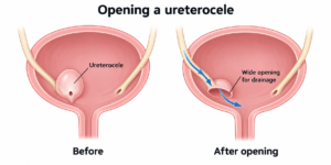
 

Information Sheet: Bladder Cancer